


 Foundational characteristics of cancer include proliferation, angiogenesis, migration, evasion of apoptosis, and cellular immortality. Find key markers for these cellular processes and antibodies to detect them.
 The SUMOplot™ Analysis Program predicts and scores sumoylation sites in your protein. SUMOylation is a post-translational modification involved in various cellular processes, such as nuclear-cytosolic transport, transcriptional regulation, apoptosis, protein stability, response to stress, and progression through the cell cycle.
 The Autophagy Receptor Motif Plotter predicts and scores autophagy receptor binding sites in your protein. Identifying proteins connected to this pathway is critical to understanding the role of autophagy in physiological as well as pathological processes such as development, differentiation, neurodegenerative diseases, stress, infection, and cancer.
Mouse Monoclonal Antibody to IDH1
Purified Mouse Monoclonal Antibody


- SPECIFICATION
- CITATIONS
- PROTOCOLS
- BACKGROUND

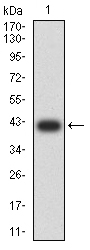
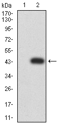
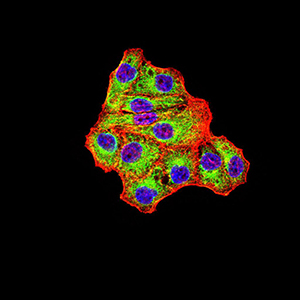
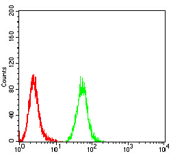
Application
| WB, FC, ICC, E |
|---|---|
| Primary Accession | O75874 |
| Reactivity | Human, Mouse, Monkey |
| Host | Mouse |
| Clonality | Monoclonal |
| Clone Names | 7G8A1 |
| Isotype | Mouse IgG1 |
| Calculated MW | 46.7kDa |
| Description | Isocitrate dehydrogenases catalyze the oxidative decarboxylation of isocitrate to 2-oxoglutarate. These enzymes belong to two distinct subclasses, one of which utilizes NAD(+) as the electron acceptor and the other NADP(+). Five isocitrate dehydrogenases have been reported: three NAD(+)-dependent isocitrate dehydrogenases, which localize to the mitochondrial matrix, and two NADP(+)-dependent isocitrate dehydrogenases, one of which is mitochondrial and the other predominantly cytosolic. Each NADP(+)-dependent isozyme is a homodimer. The protein encoded by this gene is the NADP(+)-dependent isocitrate dehydrogenase found in the cytoplasm and peroxisomes. It contains the PTS-1 peroxisomal targeting signal sequence. The presence of this enzyme in peroxisomes suggests roles in the regeneration of NADPH for intraperoxisomal reductions, such as the conversion of 2, 4-dienoyl-CoAs to 3-enoyl-CoAs, as well as in peroxisomal reactions that consume 2-oxoglutarate, namely the alpha-hydroxylation of phytanic acid. The cytoplasmic enzyme serves a significant role in cytoplasmic NADPH production. Alternatively spliced transcript variants encoding the same protein have been found for this gene.; |
| Immunogen | Purified recombinant fragment of human IDH1 (AA: 156-298) expressed in E. Coli. |
| Formulation | Purified antibody in PBS with 0.05% sodium azide |
| Application Note | ELISA: 1/10000 WB: 1/500 - 1/2000 ICC: 1/50 - 1/250 FCM: 1/200 - 1/400 |
| Gene ID | 3417 |
|---|---|
| Other Names | IDH; IDP; IDCD; IDPC; PICD; HEL-216; HEL-S-26 |
| Dilution | WB~~1:1000 FC~~1:10~50 ICC~~N/A E~~N/A |
| Storage | Maintain refrigerated at 2-8°C for up to 6 months. For long term storage store at -20°C in small aliquots to prevent freeze-thaw cycles. |
| Precautions | Mouse Monoclonal Antibody to IDH1 is for research use only and not for use in diagnostic or therapeutic procedures. |
| Name | IDH1 |
|---|---|
| Synonyms | PICD |
| Function | Catalyzes the NADP(+)-dependent oxidative decarboxylation of isocitrate (D-threo-isocitrate) to 2-ketoglutarate (2-oxoglutarate), which is required by other enzymes such as the phytanoyl-CoA dioxygenase (PubMed:10521434, PubMed:19935646). Plays a critical role in the generation of NADPH, an important cofactor in many biosynthesis pathways (PubMed:10521434). May act as a corneal epithelial crystallin and may be involved in maintaining corneal epithelial transparency (By similarity). |
| Cellular Location | Cytoplasm, cytosol. Peroxisome |

Thousands of laboratories across the world have published research that depended on the performance of antibodies from Abcepta to advance their research. Check out links to articles that cite our products in major peer-reviewed journals, organized by research category.
info@abcepta.com, and receive a free "I Love Antibodies" mug.

Provided below are standard protocols that you may find useful for product applications.
References
1.Cancer Cell. 2015 Dec 14;28(6):773-84. ; 2.Int J Cancer. 2015 Sep 1;137(5):1058-65. ;
If you have used an Abcepta product and would like to share how it has performed, please click on the "Submit Review" button and provide the requested information. Our staff will examine and post your review and contact you if needed.
If you have any additional inquiries please email technical services at tech@abcepta.com.
Ordering Information
Other Products
Shipping Information
