


 Foundational characteristics of cancer include proliferation, angiogenesis, migration, evasion of apoptosis, and cellular immortality. Find key markers for these cellular processes and antibodies to detect them.
 The SUMOplot™ Analysis Program predicts and scores sumoylation sites in your protein. SUMOylation is a post-translational modification involved in various cellular processes, such as nuclear-cytosolic transport, transcriptional regulation, apoptosis, protein stability, response to stress, and progression through the cell cycle.
 The Autophagy Receptor Motif Plotter predicts and scores autophagy receptor binding sites in your protein. Identifying proteins connected to this pathway is critical to understanding the role of autophagy in physiological as well as pathological processes such as development, differentiation, neurodegenerative diseases, stress, infection, and cancer.
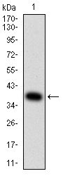
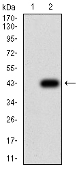
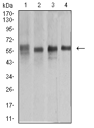
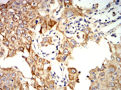
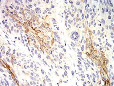
Mouse Monoclonal Antibody to KRT10
Purified Mouse Monoclonal Antibody

- SPECIFICATION
- CITATIONS
- PROTOCOLS
- BACKGROUND

Application
| WB, IHC, E |
|---|---|
| Primary Accession | P13645 |
| Reactivity | Human, Monkey |
| Host | Mouse |
| Clonality | Monoclonal |
| Clone Names | 1C3D9 |
| Isotype | Mouse IgG1 |
| Calculated MW | 58.8kDa |
| Description | This gene encodes a member of the type I (acidic) cytokeratin family, which belongs to the superfamily of intermediate filament (IF) proteins. Keratins are heteropolymeric structural proteins which form the intermediate filament. These filaments, along with actin microfilaments and microtubules, compose the cytoskeleton of epithelial cells. Mutations in this gene are associated with epidermolytic hyperkeratosis. This gene is located within a cluster of keratin family members on chromosome 17q21.; |
| Immunogen | Purified recombinant fragment of human KRT10 (AA: 345-454 ) expressed in E. Coli. |
| Formulation | Purified antibody in PBS with 0.05% sodium azide |
| Application Note | ELISA: 1/10000 WB: 1/500 - 1/2000 IHC: 1/200 - 1/1000 |
| Gene ID | 3858 |
|---|---|
| Other Names | BIE; EHK; K10; KPP; BCIE; CK10 |
| Dilution | WB~~1:1000 IHC~~1:100~500 E~~N/A |
| Storage | Maintain refrigerated at 2-8°C for up to 6 months. For long term storage store at -20°C in small aliquots to prevent freeze-thaw cycles. |
| Precautions | Mouse Monoclonal Antibody to KRT10 is for research use only and not for use in diagnostic or therapeutic procedures. |
| Name | KRT10 |
|---|---|
| Synonyms | KPP |
| Function | Plays a role in the establishment of the epidermal barrier on plantar skin (By similarity). Involved in the maintenance of cell layer development and keratin filament bundles in suprabasal cells of the epithelium (By similarity). |
| Cellular Location | Secreted, extracellular space. Cell surface. Cytoplasm |
| Tissue Location | Seen in all suprabasal cell layers including stratum corneum. Expressed on the surface of lung cell lines (PubMed:19627498). Localized on the surface of desquamated nasal epithelial cells (at protein level) (PubMed:12427098) |

Thousands of laboratories across the world have published research that depended on the performance of antibodies from Abcepta to advance their research. Check out links to articles that cite our products in major peer-reviewed journals, organized by research category.
info@abcepta.com, and receive a free "I Love Antibodies" mug.

Provided below are standard protocols that you may find useful for product applications.
References
1.JAMA Dermatol. 2015 Jan;151(1):64-9.; 2.Histopathology. 2012 Nov;61(5):910-20. ;
If you have used an Abcepta product and would like to share how it has performed, please click on the "Submit Review" button and provide the requested information. Our staff will examine and post your review and contact you if needed.
If you have any additional inquiries please email technical services at tech@abcepta.com.
Ordering Information
Other Products
Shipping Information
