


 Foundational characteristics of cancer include proliferation, angiogenesis, migration, evasion of apoptosis, and cellular immortality. Find key markers for these cellular processes and antibodies to detect them.
 The SUMOplot™ Analysis Program predicts and scores sumoylation sites in your protein. SUMOylation is a post-translational modification involved in various cellular processes, such as nuclear-cytosolic transport, transcriptional regulation, apoptosis, protein stability, response to stress, and progression through the cell cycle.
 The Autophagy Receptor Motif Plotter predicts and scores autophagy receptor binding sites in your protein. Identifying proteins connected to this pathway is critical to understanding the role of autophagy in physiological as well as pathological processes such as development, differentiation, neurodegenerative diseases, stress, infection, and cancer.
Mouse Monoclonal Antibody to ATG2A
Purified Mouse Monoclonal Antibody
- SPECIFICATION
- CITATIONS
- PROTOCOLS
- BACKGROUND

Application
| WB, ICC, E |
|---|---|
| Primary Accession | Q2TAZ0 |
| Reactivity | Human |
| Host | Mouse |
| Clonality | Monoclonal |
| Clone Names | 4H8G3 |
| Isotype | Mouse IgG1 |
| Calculated MW | 212.8kDa |
| Description | ATG2A (Autophagy Related 2A) is a Protein Coding gene. An important paralog of this gene is ATG2B.; |
| Immunogen | Purified recombinant fragment of human ATG2A (AA: 325-429) expressed in E. Coli. |
| Formulation | Purified antibody in PBS with 0.05% sodium azide |
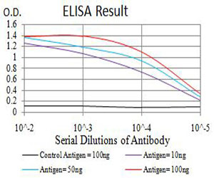
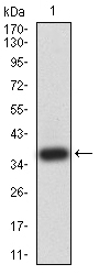
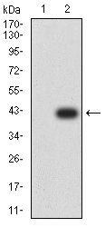
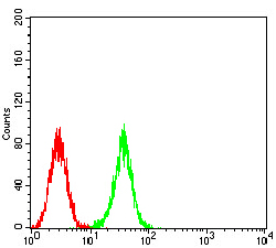
| Application Note | ELISA: 1/10000 WB: 1/500 - 1/2000 ICC: 1/200 - 1/1000 |
| Gene ID | 23130 |
|---|---|
| Dilution | WB~~1:1000 ICC~~N/A E~~N/A |
| Storage | Maintain refrigerated at 2-8°C for up to 6 months. For long term storage store at -20°C in small aliquots to prevent freeze-thaw cycles. |
| Precautions | Mouse Monoclonal Antibody to ATG2A is for research use only and not for use in diagnostic or therapeutic procedures. |
| Name | ATG2A {ECO:0000303|PubMed:21887408, ECO:0000312|HGNC:HGNC:29028} |
|---|---|
| Function | Lipid transfer protein involved in autophagosome assembly (PubMed:28561066, PubMed:30952800, PubMed:31271352). Tethers the edge of the isolation membrane (IM) to the endoplasmic reticulum (ER) and mediates direct lipid transfer from ER to IM for IM expansion (PubMed:30952800, PubMed:31271352). Binds to the ER exit site (ERES), which is the membrane source for autophagosome formation, and extracts phospholipids from the membrane source and transfers them to ATG9 (ATG9A or ATG9B) to the IM for membrane expansion (PubMed:30952800, PubMed:31271352). Lipid transfer activity is enhanced by WIPI1 and WDR45/WIPI4, which promote ATG2A-association with phosphatidylinositol 3-monophosphate (PI3P)-containing membranes (PubMed:31271352). Also regulates lipid droplets morphology and distribution within the cell (PubMed:22219374, PubMed:28561066). |
| Cellular Location | Preautophagosomal structure membrane; Peripheral membrane protein {ECO:0000250|UniProtKB:Q96BY7}. Lipid droplet {ECO:0000250|UniProtKB:Q96BY7}. Endoplasmic reticulum membrane; Peripheral membrane protein {ECO:0000250|UniProtKB:P53855}. Note=Localizes to endoplasmic reticulum-autophagosome contact sites. |

Thousands of laboratories across the world have published research that depended on the performance of antibodies from Abcepta to advance their research. Check out links to articles that cite our products in major peer-reviewed journals, organized by research category.
info@abcepta.com, and receive a free "I Love Antibodies" mug.

Provided below are standard protocols that you may find useful for product applications.
References
1.Mol Biol Cell. 2012 Mar;23(5):896-909. ; 2.Acta Biochim Pol. 2011;58(3):365-74.;
If you have used an Abcepta product and would like to share how it has performed, please click on the "Submit Review" button and provide the requested information. Our staff will examine and post your review and contact you if needed.
If you have any additional inquiries please email technical services at tech@abcepta.com.
Ordering Information
Other Products
Shipping Information
