


 Foundational characteristics of cancer include proliferation, angiogenesis, migration, evasion of apoptosis, and cellular immortality. Find key markers for these cellular processes and antibodies to detect them.
 The SUMOplot™ Analysis Program predicts and scores sumoylation sites in your protein. SUMOylation is a post-translational modification involved in various cellular processes, such as nuclear-cytosolic transport, transcriptional regulation, apoptosis, protein stability, response to stress, and progression through the cell cycle.
 The Autophagy Receptor Motif Plotter predicts and scores autophagy receptor binding sites in your protein. Identifying proteins connected to this pathway is critical to understanding the role of autophagy in physiological as well as pathological processes such as development, differentiation, neurodegenerative diseases, stress, infection, and cancer.
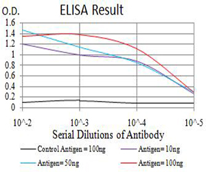
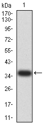
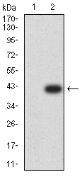
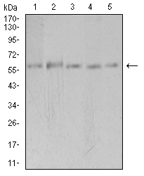
PAK3
Purified Mouse Monoclonal Antibody
- SPECIFICATION
- CITATIONS
- PROTOCOLS
- BACKGROUND

Application
| WB, IHC, ICC, E |
|---|---|
| Primary Accession | O75914 |
| Reactivity | Human, Monkey |
| Host | Mouse |
| Clonality | Monoclonal |
| Clone Names | 4G8F11 |
| Isotype | Mouse IgG1 |
| Calculated MW | 62.3kDa |
| Immunogen | Purified recombinant fragment of human PAK3 (AA: 1-100) expressed in E. Coli. |
| Formulation | Purified antibody in PBS with 0.05% sodium azide |
| Gene ID | 5063 |
|---|---|
| Other Names | ARA; bPAK; MRX30; MRX47; OPHN3; PAK-3; PAK3beta; beta-PAK |
| Dilution | WB~~ 1/500 - 1/2000 IHC~~1:100~500 ICC~~N/A E~~ 1/10000 |
| Storage | Maintain refrigerated at 2-8°C for up to 6 months. For long term storage store at -20°C in small aliquots to prevent freeze-thaw cycles. |
| Precautions | PAK3 is for research use only and not for use in diagnostic or therapeutic procedures. |
| Name | PAK3 |
|---|---|
| Synonyms | OPHN3 |
| Function | Serine/threonine protein kinase that plays a role in a variety of different signaling pathways including cytoskeleton regulation, cell migration, or cell cycle regulation. Plays a role in dendrite spine morphogenesis as well as synapse formation and plasticity. Acts as a downstream effector of the small GTPases CDC42 and RAC1. Activation by the binding of active CDC42 and RAC1 results in a conformational change and a subsequent autophosphorylation on several serine and/or threonine residues. Phosphorylates MAPK4 and MAPK6 and activates the downstream target MAPKAPK5, a regulator of F-actin polymerization and cell migration. Additionally, phosphorylates TNNI3/troponin I to modulate calcium sensitivity and relaxation kinetics of thin myofilaments. May also be involved in early neuronal development. In hippocampal neurons, necessary for the formation of dendritic spines and excitatory synapses; this function is dependent on kinase activity and may be exerted by the regulation of actomyosin contractility through the phosphorylation of myosin II regulatory light chain (MLC) (By similarity). |
| Cellular Location | Cytoplasm. |
| Tissue Location | Restricted to the nervous system. Highly expressed in postmitotic neurons of the developing and postnatal cerebral cortex and hippocampus. |

Thousands of laboratories across the world have published research that depended on the performance of antibodies from Abcepta to advance their research. Check out links to articles that cite our products in major peer-reviewed journals, organized by research category.
info@abcepta.com, and receive a free "I Love Antibodies" mug.

Provided below are standard protocols that you may find useful for product applications.
References
1.J Mol Biol. 2014 Oct 23;426(21):3520-38.2.J Biol Chem. 2011 Nov 18;286(46):40044-59.
If you have used an Abcepta product and would like to share how it has performed, please click on the "Submit Review" button and provide the requested information. Our staff will examine and post your review and contact you if needed.
If you have any additional inquiries please email technical services at tech@abcepta.com.
Ordering Information
Other Products
Shipping Information
