


 Foundational characteristics of cancer include proliferation, angiogenesis, migration, evasion of apoptosis, and cellular immortality. Find key markers for these cellular processes and antibodies to detect them.
 The SUMOplot™ Analysis Program predicts and scores sumoylation sites in your protein. SUMOylation is a post-translational modification involved in various cellular processes, such as nuclear-cytosolic transport, transcriptional regulation, apoptosis, protein stability, response to stress, and progression through the cell cycle.
 The Autophagy Receptor Motif Plotter predicts and scores autophagy receptor binding sites in your protein. Identifying proteins connected to this pathway is critical to understanding the role of autophagy in physiological as well as pathological processes such as development, differentiation, neurodegenerative diseases, stress, infection, and cancer.
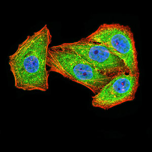
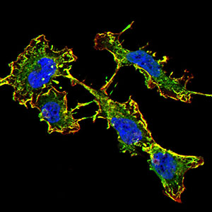
GRIN2B
Purified Mouse Monoclonal Antibody
- SPECIFICATION
- CITATIONS
- PROTOCOLS
- BACKGROUND

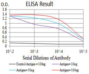
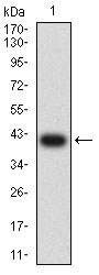
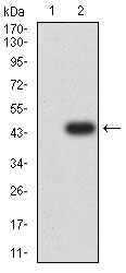
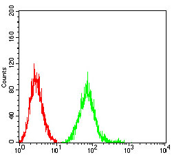
Application
| WB, IHC, ICC, E |
|---|---|
| Primary Accession | Q13224 |
| Reactivity | Human |
| Host | Mouse |
| Clonality | Monoclonal |
| Clone Names | 6E9A8 |
| Isotype | Mouse IgG1 |
| Calculated MW | 166.4kDa |
| Immunogen | Purified recombinant fragment of human GRIN2B (AA: extra 27-163) expressed in E. Coli. |
| Formulation | Purified antibody in PBS with 0.05% sodium azide |
| Gene ID | 2904 |
|---|---|
| Other Names | MRD6; NR2B; hNR3; EIEE27; GluN2B; NMDAR2B |
| Dilution | WB~~ 1/500 - 1/2000 IHC~~1:100~500 ICC~~ 1/100 - 1/500 E~~ 1/10000 |
| Storage | Maintain refrigerated at 2-8°C for up to 6 months. For long term storage store at -20°C in small aliquots to prevent freeze-thaw cycles. |
| Precautions | GRIN2B is for research use only and not for use in diagnostic or therapeutic procedures. |
| Name | GRIN2B {ECO:0000303|Ref.3, ECO:0000312|HGNC:HGNC:4586} |
|---|---|
| Function | Component of N-methyl-D-aspartate (NMDA) receptors (NMDARs) that function as heterotetrameric, ligand-gated cation channels with high calcium permeability and voltage-dependent block by Mg(2+) (PubMed:24272827, PubMed:24863970, PubMed:26875626, PubMed:26919761, PubMed:27839871, PubMed:28095420, PubMed:28126851, PubMed:38538865, PubMed:8768735). Participates in synaptic plasticity for learning and memory formation by contributing to the long-term depression (LTD) of hippocampus membrane currents (By similarity). Channel activation requires binding of the neurotransmitter L-glutamate to the GluN2 subunit, glycine or D-serine binding to the GluN1 subunit, plus membrane depolarization to eliminate channel inhibition by Mg(2+) (PubMed:24272827, PubMed:24863970, PubMed:26875626, PubMed:26919761, PubMed:27839871, PubMed:28095420, PubMed:28126851, PubMed:38538865, PubMed:8768735). NMDARs mediate simultaneously the potasium efflux and the influx of calcium and sodium (By similarity). Each GluN2 subunit confers differential attributes to channel properties, including activation, deactivation and desensitization kinetics, pH sensitivity, Ca2(+) permeability, and binding to allosteric modulators (PubMed:26875626, PubMed:28095420, PubMed:28126851, PubMed:38538865, PubMed:8768735). In concert with DAPK1 at extrasynaptic sites, acts as a central mediator for stroke damage. Its phosphorylation at Ser-1303 by DAPK1 enhances synaptic NMDA receptor channel activity inducing injurious Ca2+ influx through them, resulting in an irreversible neuronal death (By similarity). |
| Cellular Location | Cell membrane; Multi-pass membrane protein. Postsynaptic cell membrane {ECO:0000250|UniProtKB:Q00960}; Multi-pass membrane protein. Cell projection, dendrite. Late endosome {ECO:0000250|UniProtKB:Q01097}. Lysosome {ECO:0000250|UniProtKB:Q01097}. Cytoplasm, cytoskeleton {ECO:0000250|UniProtKB:Q01097}. Note=Co-localizes with the motor protein KIF17 along microtubules. {ECO:0000250|UniProtKB:Q01097} |
| Tissue Location | Primarily found in the fronto-parieto-temporal cortex and hippocampus pyramidal cells, lower expression in the basal ganglia. |

Thousands of laboratories across the world have published research that depended on the performance of antibodies from Abcepta to advance their research. Check out links to articles that cite our products in major peer-reviewed journals, organized by research category.
info@abcepta.com, and receive a free "I Love Antibodies" mug.

Provided below are standard protocols that you may find useful for product applications.
References
1.J Neural Transm (Vienna). 2014 May;121(5):533-42. 2.Psychopharmacology (Berl). 2014 Feb;231(4):685-93.
If you have used an Abcepta product and would like to share how it has performed, please click on the "Submit Review" button and provide the requested information. Our staff will examine and post your review and contact you if needed.
If you have any additional inquiries please email technical services at tech@abcepta.com.
Ordering Information
Other Products
Shipping Information
