


 Foundational characteristics of cancer include proliferation, angiogenesis, migration, evasion of apoptosis, and cellular immortality. Find key markers for these cellular processes and antibodies to detect them.
 The SUMOplot™ Analysis Program predicts and scores sumoylation sites in your protein. SUMOylation is a post-translational modification involved in various cellular processes, such as nuclear-cytosolic transport, transcriptional regulation, apoptosis, protein stability, response to stress, and progression through the cell cycle.
 The Autophagy Receptor Motif Plotter predicts and scores autophagy receptor binding sites in your protein. Identifying proteins connected to this pathway is critical to understanding the role of autophagy in physiological as well as pathological processes such as development, differentiation, neurodegenerative diseases, stress, infection, and cancer.
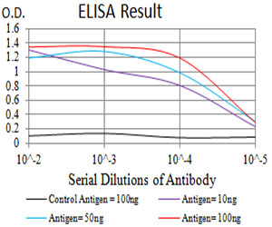
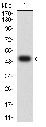
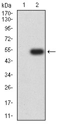
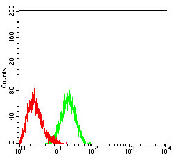
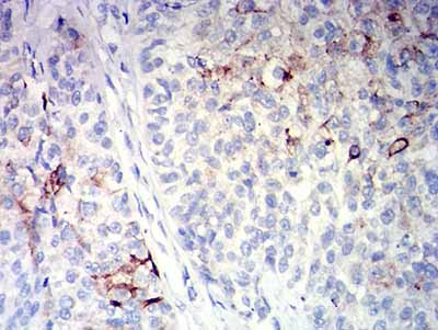
CD10
Purified Mouse Monoclonal Antibody
- SPECIFICATION
- CITATIONS
- PROTOCOLS
- BACKGROUND

Application
| WB, IHC, ICC, E |
|---|---|
| Primary Accession | P08473 |
| Reactivity | Human |
| Host | Mouse |
| Clonality | Monoclonal |
| Clone Names | 3G6B11 |
| Isotype | Mouse IgG1 |
| Calculated MW | 85.5kDa |
| Immunogen | Purified recombinant fragment of human CD10 (AA: extra 549-750) expressed in E. Coli. |
| Formulation | Purified antibody in PBS with 0.05% sodium azide |
| Gene ID | 4311 |
|---|---|
| Other Names | NEP; SFE; MME; CALLA; CMT2T; SCA43 |
| Dilution | WB~~ 1/500 - 1/2000 IHC~~1/200 - 1/1000 ICC~~N/A E~~ 1/10000 |
| Storage | Maintain refrigerated at 2-8°C for up to 6 months. For long term storage store at -20°C in small aliquots to prevent freeze-thaw cycles. |
| Precautions | CD10 is for research use only and not for use in diagnostic or therapeutic procedures. |
| Name | MME {ECO:0000303|PubMed:27588448, ECO:0000312|HGNC:HGNC:7154} |
|---|---|
| Function | Thermolysin-like specificity, but is almost confined on acting on polypeptides of up to 30 amino acids (PubMed:15283675, PubMed:6208535, PubMed:6349683, PubMed:8168535). Biologically important in the destruction of opioid peptides such as Met- and Leu-enkephalins by cleavage of a Gly-Phe bond (PubMed:17101991, PubMed:6349683). Catalyzes cleavage of bradykinin, substance P and neurotensin peptides (PubMed:6208535). Able to cleave angiotensin-1, angiotensin-2 and angiotensin 1-9 (PubMed:15283675, PubMed:6349683). Involved in the degradation of atrial natriuretic factor (ANF) and brain natriuretic factor (BNP(1-32)) (PubMed:16254193, PubMed:2531377, PubMed:2972276). Displays UV-inducible elastase activity toward skin preelastic and elastic fibers (PubMed:20876573). |
| Cellular Location | Cell membrane; Single-pass type II membrane protein |

Thousands of laboratories across the world have published research that depended on the performance of antibodies from Abcepta to advance their research. Check out links to articles that cite our products in major peer-reviewed journals, organized by research category.
info@abcepta.com, and receive a free "I Love Antibodies" mug.

Provided below are standard protocols that you may find useful for product applications.
References
1.Pathobiology. 2015;82(6):259-63. 2.Asian Pac J Cancer Prev. 2015;16(8):3147-52.
If you have used an Abcepta product and would like to share how it has performed, please click on the "Submit Review" button and provide the requested information. Our staff will examine and post your review and contact you if needed.
If you have any additional inquiries please email technical services at tech@abcepta.com.
Ordering Information
Other Products
Shipping Information
