


 Foundational characteristics of cancer include proliferation, angiogenesis, migration, evasion of apoptosis, and cellular immortality. Find key markers for these cellular processes and antibodies to detect them.
 The SUMOplot™ Analysis Program predicts and scores sumoylation sites in your protein. SUMOylation is a post-translational modification involved in various cellular processes, such as nuclear-cytosolic transport, transcriptional regulation, apoptosis, protein stability, response to stress, and progression through the cell cycle.
 The Autophagy Receptor Motif Plotter predicts and scores autophagy receptor binding sites in your protein. Identifying proteins connected to this pathway is critical to understanding the role of autophagy in physiological as well as pathological processes such as development, differentiation, neurodegenerative diseases, stress, infection, and cancer.
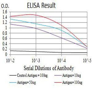
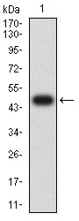
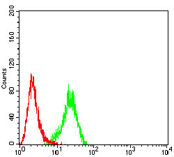
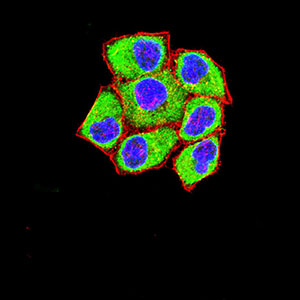
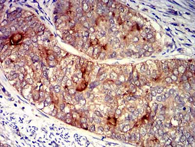
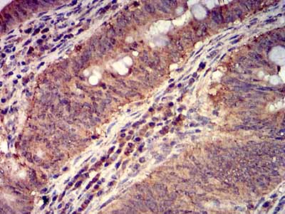
FOLR1
Purified Mouse Monoclonal Antibody

- SPECIFICATION
- CITATIONS
- PROTOCOLS
- BACKGROUND

Application
| WB, IHC, ICC, E |
|---|---|
| Primary Accession | P15328 |
| Reactivity | Human |
| Host | Mouse |
| Clonality | Monoclonal |
| Clone Names | 2G5C12 |
| Isotype | Mouse IgG2a |
| Calculated MW | 29.8kDa |
| Immunogen | Purified recombinant fragment of human FOLR1 (AA: 41-227) expressed in E. Coli. |
| Formulation | Purified antibody in PBS with 0.05% sodium azide |
| Gene ID | 2348 |
|---|---|
| Other Names | FBP; FOLR |
| Dilution | WB~~ 1/500 - 1/2000 IHC~~ 1/200 - 1/1000 ICC~~ 1/100 - 1/500 E~~ 1/10000 |
| Storage | Maintain refrigerated at 2-8°C for up to 6 months. For long term storage store at -20°C in small aliquots to prevent freeze-thaw cycles. |
| Precautions | FOLR1 is for research use only and not for use in diagnostic or therapeutic procedures. |
| Name | FOLR1 |
|---|---|
| Synonyms | FOLR |
| Function | Binds to folate and reduced folic acid derivatives and mediates delivery of 5-methyltetrahydrofolate and folate analogs into the interior of cells (PubMed:19074442, PubMed:23851396, PubMed:23934049, PubMed:2527252, PubMed:8033114, PubMed:8567728). Has high affinity for folate and folic acid analogs at neutral pH (PubMed:23851396, PubMed:23934049, PubMed:2527252, PubMed:8033114, PubMed:8567728). Exposure to slightly acidic pH after receptor endocytosis triggers a conformation change that strongly reduces its affinity for folates and mediates their release (PubMed:8567728). Required for normal embryonic development and normal cell proliferation (By similarity). |
| Cellular Location | Cell membrane; Lipid-anchor, GPI-anchor Apical cell membrane; Lipid-anchor, GPI- anchor Basolateral cell membrane; Lipid-anchor, GPI-like-anchor. Secreted Cytoplasmic vesicle. Cytoplasmic vesicle, clathrin-coated vesicle. Endosome. Note=Endocytosed into cytoplasmic vesicles and then recycled to the cell membrane |
| Tissue Location | Primarily expressed in tissues of epithelial origin. Expression is increased in malignant tissues. Expressed in kidney, lung and cerebellum. Detected in placenta and thymus epithelium. |

Thousands of laboratories across the world have published research that depended on the performance of antibodies from Abcepta to advance their research. Check out links to articles that cite our products in major peer-reviewed journals, organized by research category.
info@abcepta.com, and receive a free "I Love Antibodies" mug.

Provided below are standard protocols that you may find useful for product applications.
References
1.Biosens Bioelectron. 2016 Apr 15;78:147-53.2.PLoS One. 2015 Mar 27;10(3):e0122209.
If you have used an Abcepta product and would like to share how it has performed, please click on the "Submit Review" button and provide the requested information. Our staff will examine and post your review and contact you if needed.
If you have any additional inquiries please email technical services at tech@abcepta.com.
Ordering Information
Other Products
Shipping Information
