


 Foundational characteristics of cancer include proliferation, angiogenesis, migration, evasion of apoptosis, and cellular immortality. Find key markers for these cellular processes and antibodies to detect them.
 The SUMOplot™ Analysis Program predicts and scores sumoylation sites in your protein. SUMOylation is a post-translational modification involved in various cellular processes, such as nuclear-cytosolic transport, transcriptional regulation, apoptosis, protein stability, response to stress, and progression through the cell cycle.
 The Autophagy Receptor Motif Plotter predicts and scores autophagy receptor binding sites in your protein. Identifying proteins connected to this pathway is critical to understanding the role of autophagy in physiological as well as pathological processes such as development, differentiation, neurodegenerative diseases, stress, infection, and cancer.
INPP5B Antibody (C-term)
Affinity Purified Rabbit Polyclonal Antibody (Pab)
- SPECIFICATION
- CITATIONS
- PROTOCOLS
- BACKGROUND

Application
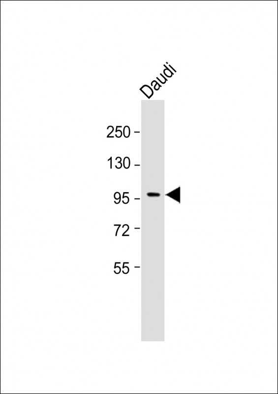
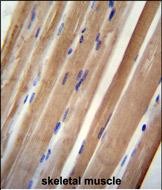
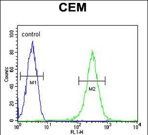
| WB, FC, IHC-P, E |
|---|---|
| Primary Accession | P32019 |
| Other Accession | NP_005531.2 |
| Reactivity | Human |
| Host | Rabbit |
| Clonality | Polyclonal |
| Isotype | Rabbit IgG |
| Calculated MW | 112852 Da |
| Antigen Region | 950-979 aa |
| Gene ID | 3633 |
|---|---|
| Other Names | Type II inositol 1, 5-trisphosphate 5-phosphatase, 75 kDa inositol polyphosphate-5-phosphatase, Phosphoinositide 5-phosphatase, 5PTase, INPP5B, OCRL2 |
| Target/Specificity | This INPP5B antibody is generated from rabbits immunized with a KLH conjugated synthetic peptide between 950-979 amino acids from the C-terminal region of human INPP5B. |
| Dilution | WB~~1:1000 FC~~1:10~50 IHC-P~~1:50~100 E~~Use at an assay dependent concentration. |
| Format | Purified polyclonal antibody supplied in PBS with 0.09% (W/V) sodium azide. This antibody is purified through a protein A column, followed by peptide affinity purification. |
| Storage | Maintain refrigerated at 2-8°C for up to 2 weeks. For long term storage store at -20°C in small aliquots to prevent freeze-thaw cycles. |
| Precautions | INPP5B Antibody (C-term) is for research use only and not for use in diagnostic or therapeutic procedures. |
| Name | INPP5B |
|---|---|
| Synonyms | OCRL2 |
| Function | Hydrolyzes phosphatidylinositol 4,5-bisphosphate (PtIns(4,5)P2) and the signaling molecule phosphatidylinositol 1,4,5- trisphosphate (PtIns(1,4,5)P3), and thereby modulates cellular signaling events. |
| Cellular Location | Cytoplasm, cytosol. Endoplasmic reticulum-Golgi intermediate compartment. Early endosome membrane. Membrane; Peripheral membrane protein; Cytoplasmic side. Cytoplasmic vesicle, phagosome membrane {ECO:0000250|UniProtKB:Q8K337}. Golgi apparatus |
| Tissue Location | Platelets. |

Thousands of laboratories across the world have published research that depended on the performance of antibodies from Abcepta to advance their research. Check out links to articles that cite our products in major peer-reviewed journals, organized by research category.
info@abcepta.com, and receive a free "I Love Antibodies" mug.

Provided below are standard protocols that you may find useful for product applications.
Background
Cellular calcium signaling is controlled by the production of inositol phosphates (IPs) by phospholipase C in response to extracellular signals. The IP signaling molecules are inactivated by a family of inositol polyphosphate-5-phosphatases (5-phosphatases). INPP5B encodes the type II 5-phosphatase. The protein is localized to the cytosol and mitochondria, and associates with membranes through an isoprenyl modification near the C-terminus. Several alternatively spliced transcript variants of this gene have been described, but the full-length nature of some of these variants has not been determined. [provided by RefSeq].
References
Coon, B.G., et al. Hum. Mol. Genet. 18(23):4478-4491(2009)
Mao, Y., et al. EMBO J. 28(13):1831-1842(2009)
Williams, C., et al. J. Cell. Sci. 120 (PT 22), 3941-3951 (2007) :
Speed, C.J., et al. Eur. J. Biochem. 234(1):216-224(1995)
Janne, P.A., et al. Genomics 28(2):280-285(1995)
If you have used an Abcepta product and would like to share how it has performed, please click on the "Submit Review" button and provide the requested information. Our staff will examine and post your review and contact you if needed.
If you have any additional inquiries please email technical services at tech@abcepta.com.
Ordering Information
Other Products
Shipping Information
