


 Foundational characteristics of cancer include proliferation, angiogenesis, migration, evasion of apoptosis, and cellular immortality. Find key markers for these cellular processes and antibodies to detect them.
 The SUMOplot™ Analysis Program predicts and scores sumoylation sites in your protein. SUMOylation is a post-translational modification involved in various cellular processes, such as nuclear-cytosolic transport, transcriptional regulation, apoptosis, protein stability, response to stress, and progression through the cell cycle.
 The Autophagy Receptor Motif Plotter predicts and scores autophagy receptor binding sites in your protein. Identifying proteins connected to this pathway is critical to understanding the role of autophagy in physiological as well as pathological processes such as development, differentiation, neurodegenerative diseases, stress, infection, and cancer.
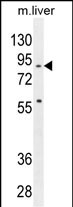
SPIRE2 Antibody (Center)
Affinity Purified Rabbit Polyclonal Antibody (Pab)
- SPECIFICATION
- CITATIONS
- PROTOCOLS
- BACKGROUND

Application
| WB, E |
|---|---|
| Primary Accession | Q8WWL2 |
| Other Accession | Q8K1S6, NP_115827.1 |
| Reactivity | Human, Mouse |
| Host | Rabbit |
| Clonality | Polyclonal |
| Isotype | Rabbit IgG |
| Calculated MW | 79671 Da |
| Antigen Region | 285-313 aa |
| Gene ID | 84501 |
|---|---|
| Other Names | Protein spire homolog 2, Spir-2, SPIRE2, KIAA1832, SPIR2 |
| Target/Specificity | This SPIRE2 antibody is generated from rabbits immunized with a KLH conjugated synthetic peptide between 285-313 amino acids from the Central region of human SPIRE2. |
| Dilution | WB~~1:1000 E~~Use at an assay dependent concentration. |
| Format | Purified polyclonal antibody supplied in PBS with 0.09% (W/V) sodium azide. This antibody is purified through a protein A column, followed by peptide affinity purification. |
| Storage | Maintain refrigerated at 2-8°C for up to 2 weeks. For long term storage store at -20°C in small aliquots to prevent freeze-thaw cycles. |
| Precautions | SPIRE2 Antibody (Center) is for research use only and not for use in diagnostic or therapeutic procedures. |
| Name | SPIRE2 |
|---|---|
| Synonyms | KIAA1832, SPIR2 |
| Function | Acts as an actin nucleation factor, remains associated with the slow-growing pointed end of the new filament (PubMed:21620703). Involved in intracellular vesicle transport along actin fibers, providing a novel link between actin cytoskeleton dynamics and intracellular transport (By similarity). Required for asymmetric spindle positioning and asymmetric cell division during meiosis (PubMed:21620703). Required for normal formation of the cleavage furrow and for polar body extrusion during female germ cell meiosis (PubMed:21620703). Also acts in the nucleus: together with SPIRE1 and SPIRE2, promotes assembly of nuclear actin filaments in response to DNA damage in order to facilitate movement of chromatin and repair factors after DNA damage (PubMed:26287480). |
| Cellular Location | Cytoplasm, cytoskeleton {ECO:0000250|UniProtKB:Q8K1S6}. Cytoplasm, cytosol {ECO:0000250|UniProtKB:Q8K1S6}. Cell membrane {ECO:0000250|UniProtKB:Q8K1S6}; Peripheral membrane protein {ECO:0000250|UniProtKB:Q8K1S6}; Cytoplasmic side {ECO:0000250|UniProtKB:Q8K1S6}. Cytoplasmic vesicle membrane {ECO:0000250|UniProtKB:Q8K1S6}; Peripheral membrane protein {ECO:0000250|UniProtKB:Q8K1S6}; Cytoplasmic side {ECO:0000250|UniProtKB:Q8K1S6}. Note=Detected at the cleavage furrow during asymmetric oocyte division and polar body extrusion {ECO:0000250|UniProtKB:Q8K1S6} |

Thousands of laboratories across the world have published research that depended on the performance of antibodies from Abcepta to advance their research. Check out links to articles that cite our products in major peer-reviewed journals, organized by research category.
info@abcepta.com, and receive a free "I Love Antibodies" mug.

Provided below are standard protocols that you may find useful for product applications.
Background
Acts as a actin nucleation factor, remains associated with the slow-growing pointed end of the new filament. Involved in vesicle transport processes providing a novel link between actin organization and intracellular transport (By similarity).
References
Pechlivanis, M., et al. J. Biol. Chem. 284(37):25324-25333(2009)
If you have used an Abcepta product and would like to share how it has performed, please click on the "Submit Review" button and provide the requested information. Our staff will examine and post your review and contact you if needed.
If you have any additional inquiries please email technical services at tech@abcepta.com.
Ordering Information
Other Products
Shipping Information
