


 Foundational characteristics of cancer include proliferation, angiogenesis, migration, evasion of apoptosis, and cellular immortality. Find key markers for these cellular processes and antibodies to detect them.
 The SUMOplot™ Analysis Program predicts and scores sumoylation sites in your protein. SUMOylation is a post-translational modification involved in various cellular processes, such as nuclear-cytosolic transport, transcriptional regulation, apoptosis, protein stability, response to stress, and progression through the cell cycle.
 The Autophagy Receptor Motif Plotter predicts and scores autophagy receptor binding sites in your protein. Identifying proteins connected to this pathway is critical to understanding the role of autophagy in physiological as well as pathological processes such as development, differentiation, neurodegenerative diseases, stress, infection, and cancer.
> home > Products > Primary Antibodies > Antibody Collections > Catalog Updated > HHLA2 Antibody (N-term)
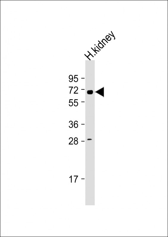
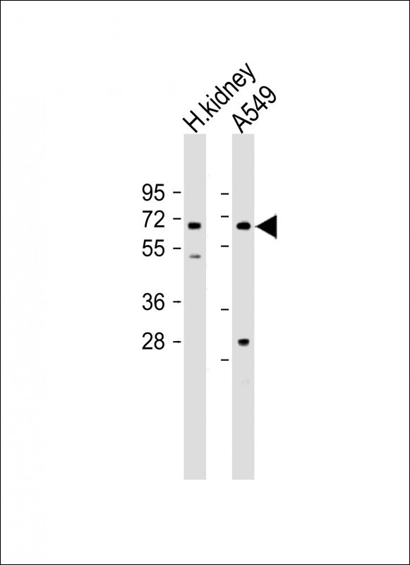
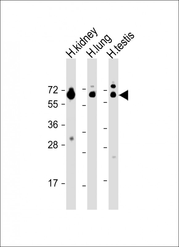
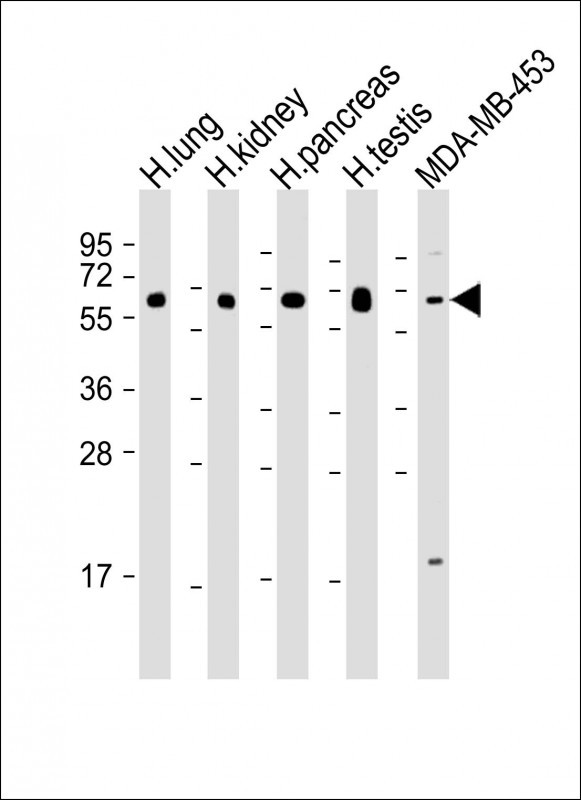
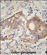
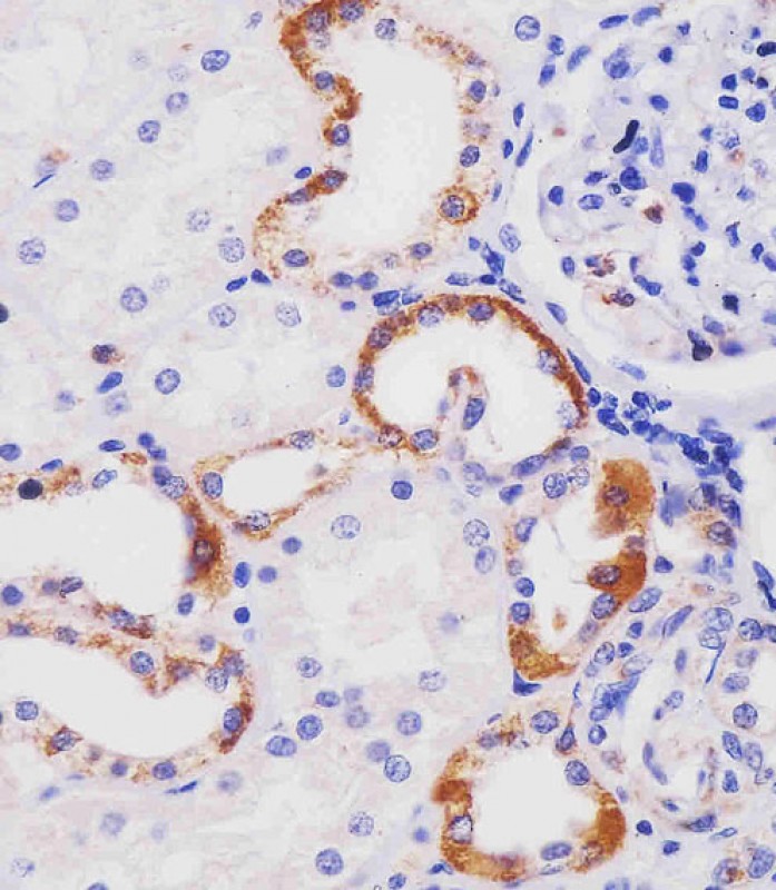
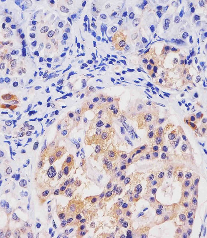
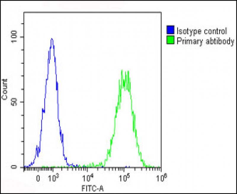
HHLA2 Antibody (N-term)
Affinity Purified Rabbit Polyclonal Antibody (Pab)
- SPECIFICATION
- CITATIONS: 1
- PROTOCOLS
- BACKGROUND

Application
| WB, IHC-P, FC, E |
|---|---|
| Primary Accession | Q9UM44 |
| Other Accession | NP_009003.1 |
| Reactivity | Human |
| Host | Rabbit |
| Clonality | Polyclonal |
| Isotype | Rabbit IgG |
| Calculated MW | 46850 Da |
| Antigen Region | 58-87 aa |
| Gene ID | 11148 |
|---|---|
| Other Names | HERV-H LTR-associating protein 2, Human endogenous retrovirus-H long terminal repeat-associating protein 2, HHLA2 |
| Target/Specificity | This HHLA2 antibody is generated from rabbits immunized with a KLH conjugated synthetic peptide between 58-87 amino acids of human HHLA2. |
| Dilution | WB~~1:2000 IHC-P~~1:25 FC~~1:25 E~~Use at an assay dependent concentration. |
| Format | Purified polyclonal antibody supplied in PBS with 0.09% (W/V) sodium azide. This antibody is purified through a protein A column, followed by peptide affinity purification. |
| Storage | Maintain refrigerated at 2-8°C for up to 2 weeks. For long term storage store at -20°C in small aliquots to prevent freeze-thaw cycles. |
| Precautions | HHLA2 Antibody (N-term) is for research use only and not for use in diagnostic or therapeutic procedures. |
| Name | HHLA2 |
|---|---|
| Function | Through interaction with TMIGD2, costimulates T-cells in the context of TCR-mediated activation. Enhances T-cell proliferation and cytokine production via an AKT-dependent signaling cascade. |
| Cellular Location | Membrane; Single-pass type I membrane protein |
| Tissue Location | Expressed at high levels in colon, kidney, testis, lung and pancreas, and at lower levels in small intestine, liver and skeletal muscle. In immune cells, highly expressed in B-cells, dendritic cells and macrophages. Not detected in T-cells |
Research Areas
Citations ( 0 )

Application Protocols
Provided below are standard protocols that you may find useful for product applications.
References
Mager, D.L., et al. Genomics 59(3):255-263(1999)
Abcepta welcomes feedback from its customers.
If you have used an Abcepta product and would like to share how it has performed, please click on the "Submit Review" button and provide the requested information. Our staff will examine and post your review and contact you if needed.
If you have any additional inquiries please email technical services at tech@abcepta.com.
$ 150.00
$ 385.00
Cat# AP10650a
Ordering Information
United States
AlbaniaAustraliaAustriaBelgiumBosnia & HerzegovinaBrazilBulgariaCanadaCentral AmericaChinaCroatiaCyprusCzech RepublicDenmarkEstoniaFinlandFranceGermanyGreeceHong KongHungaryIcelandIndiaIndonesiaIrelandIsraelItalyJapanLatviaLithuaniaLuxembourgMacedoniaMalaysiaMaltaMexicoNetherlandsNew ZealandNorwayPakistanPolandPortugalRomaniaSerbiaSingaporeSlovakiaSloveniaSouth AfricaSouth KoreaSpainSwedenSwitzerlandTaiwanTurkeyUnited KingdomUnited StatesVietnamWorldwideOthers
USA Headquarters
(888) 735-7227 / (858) 622-0099 or (858) 875-1900
Other Products
Shipping Information
Domestic orders (in stock items)
Shipped out the same day. Orders placed after 1 PM (PST) will ship out the next business day.
International orders
Contact your local distributors
