


 Foundational characteristics of cancer include proliferation, angiogenesis, migration, evasion of apoptosis, and cellular immortality. Find key markers for these cellular processes and antibodies to detect them.
 The SUMOplot™ Analysis Program predicts and scores sumoylation sites in your protein. SUMOylation is a post-translational modification involved in various cellular processes, such as nuclear-cytosolic transport, transcriptional regulation, apoptosis, protein stability, response to stress, and progression through the cell cycle.
 The Autophagy Receptor Motif Plotter predicts and scores autophagy receptor binding sites in your protein. Identifying proteins connected to this pathway is critical to understanding the role of autophagy in physiological as well as pathological processes such as development, differentiation, neurodegenerative diseases, stress, infection, and cancer.
UPF2 Antibody (Center)
Affinity Purified Rabbit Polyclonal Antibody (Pab)
- SPECIFICATION
- CITATIONS
- PROTOCOLS
- BACKGROUND

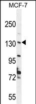
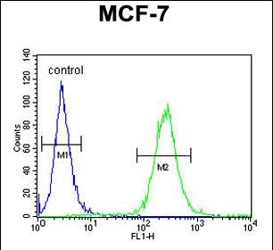
Application
| FC, WB, E |
|---|---|
| Primary Accession | Q9HAU5 |
| Other Accession | NP_056357.1 |
| Reactivity | Human |
| Host | Rabbit |
| Clonality | Polyclonal |
| Isotype | Rabbit IgG |
| Calculated MW | 147810 Da |
| Antigen Region | 630-656 aa |
| Gene ID | 26019 |
|---|---|
| Other Names | Regulator of nonsense transcripts 2, Nonsense mRNA reducing factor 2, Up-frameshift suppressor 2 homolog, hUpf2, UPF2, KIAA1408, RENT2 |
| Target/Specificity | This UPF2 antibody is generated from rabbits immunized with a KLH conjugated synthetic peptide between 630-656 amino acids from the Central region of human UPF2. |
| Dilution | FC~~1:10~50 WB~~1:1000 E~~Use at an assay dependent concentration. |
| Format | Purified polyclonal antibody supplied in PBS with 0.09% (W/V) sodium azide. This antibody is purified through a protein A column, followed by peptide affinity purification. |
| Storage | Maintain refrigerated at 2-8°C for up to 2 weeks. For long term storage store at -20°C in small aliquots to prevent freeze-thaw cycles. |
| Precautions | UPF2 Antibody (Center) is for research use only and not for use in diagnostic or therapeutic procedures. |
| Name | UPF2 (HGNC:17854) |
|---|---|
| Function | Involved in nonsense-mediated decay (NMD) of mRNAs containing premature stop codons by associating with the nuclear exon junction complex (EJC). Recruited by UPF3B associated with the EJC core at the cytoplasmic side of the nuclear envelope and the subsequent formation of an UPF1-UPF2-UPF3 surveillance complex (including UPF1 bound to release factors at the stalled ribosome) is believed to activate NMD. In cooperation with UPF3B stimulates both ATPase and RNA helicase activities of UPF1. Binds spliced mRNA. |
| Cellular Location | Cytoplasm, perinuclear region. Cytoplasm {ECO:0000250|UniProtKB:A2AT37} |
| Tissue Location | Ubiquitous.. |

Thousands of laboratories across the world have published research that depended on the performance of antibodies from Abcepta to advance their research. Check out links to articles that cite our products in major peer-reviewed journals, organized by research category.
info@abcepta.com, and receive a free "I Love Antibodies" mug.

Provided below are standard protocols that you may find useful for product applications.
Background
UPF2 is a protein that is part of a post-splicing multiprotein complex involved in both mRNA nuclear export and mRNA surveillance. mRNA surveillance detects exported mRNAs with truncated open reading frames and initiates nonsense-mediated mRNA decay (NMD). When translation ends upstream from the last exon-exon junction, this triggers NMD to degrade mRNAs containing premature stop codons. This protein is located in the perinuclear area. It interacts with translation release factors and the proteins that are functional homologs of yeast Upf1p and Upf3p.
References
Clerici, M., et al. EMBO J. 28(15):2293-2306(2009)
Cronin, S., et al. Eur. J. Hum. Genet. 17(2):213-218(2009)
Woeller, C.F., et al. EMBO Rep. 9(5):446-451(2008)
Chamieh, H., et al. Nat. Struct. Mol. Biol. 15(1):85-93(2008)
Singh, G., et al. Mol. Cell 27(5):780-792(2007)
If you have used an Abcepta product and would like to share how it has performed, please click on the "Submit Review" button and provide the requested information. Our staff will examine and post your review and contact you if needed.
If you have any additional inquiries please email technical services at tech@abcepta.com.
Ordering Information
Other Products
Shipping Information
