


 Foundational characteristics of cancer include proliferation, angiogenesis, migration, evasion of apoptosis, and cellular immortality. Find key markers for these cellular processes and antibodies to detect them.
 The SUMOplot™ Analysis Program predicts and scores sumoylation sites in your protein. SUMOylation is a post-translational modification involved in various cellular processes, such as nuclear-cytosolic transport, transcriptional regulation, apoptosis, protein stability, response to stress, and progression through the cell cycle.
 The Autophagy Receptor Motif Plotter predicts and scores autophagy receptor binding sites in your protein. Identifying proteins connected to this pathway is critical to understanding the role of autophagy in physiological as well as pathological processes such as development, differentiation, neurodegenerative diseases, stress, infection, and cancer.
CHST2 Antibody (Center)
Affinity Purified Rabbit Polyclonal Antibody (Pab)
- SPECIFICATION
- CITATIONS
- PROTOCOLS
- BACKGROUND

Application
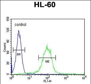
| FC, WB, E |
|---|---|
| Primary Accession | Q9Y4C5 |
| Other Accession | Q80WV3, NP_004258.2 |
| Reactivity | Human |
| Predicted | Mouse |
| Host | Rabbit |
| Clonality | Polyclonal |
| Isotype | Rabbit IgG |
| Calculated MW | 57857 Da |
| Antigen Region | 309-335 aa |
| Gene ID | 9435 |
|---|---|
| Other Names | Carbohydrate sulfotransferase 2, 282-, Galactose/N-acetylglucosamine/N-acetylglucosamine 6-O-sulfotransferase 2, GST-2, N-acetylglucosamine 6-O-sulfotransferase 1, GlcNAc6ST-1, Gn6ST-1, CHST2, GN6ST |
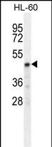
| Target/Specificity | This CHST2 antibody is generated from rabbits immunized with a KLH conjugated synthetic peptide between 309-335 amino acids from the Central region of human CHST2. |
| Dilution | FC~~1:10~50 WB~~1:1000 E~~Use at an assay dependent concentration. |
| Format | Purified polyclonal antibody supplied in PBS with 0.09% (W/V) sodium azide. This antibody is purified through a protein A column, followed by peptide affinity purification. |
| Storage | Maintain refrigerated at 2-8°C for up to 2 weeks. For long term storage store at -20°C in small aliquots to prevent freeze-thaw cycles. |
| Precautions | CHST2 Antibody (Center) is for research use only and not for use in diagnostic or therapeutic procedures. |
| Name | CHST2 |
|---|---|
| Synonyms | GN6ST |
| Function | Sulfotransferase that utilizes 3'-phospho-5'-adenylyl sulfate (PAPS) as sulfonate donor to catalyze the transfer of sulfate to position 6 of non-reducing N-acetylglucosamine (GlcNAc) residues within keratan-like structures on N- and O-linked glycans and within O-linked mucin-type glycans (PubMed:11042394, PubMed:11726653, PubMed:35939855, PubMed:38034954, PubMed:9722682). Selectively transfers the sulfate group onto the terminal GlcNAc of alpha1,3-Man or alpha1,6-Man antenna of complex-type N-glycans depending on glycan composition. Only sulfates terminal GlcNAc of alpha1,3-Man antenna of G0 complex-type N- glycans. Can sulfate keratan-type N-acetyllactosamine (LacNAc) repeats generating epitopes for self versus non-self immune recognition by C- type lectins (PubMed:35939855, PubMed:38034954). Transfers the sulfate group primarily on core 2 GlcNAcbeta1-6(Galbeta1-3)GalNAcalpha-Ser/Thr and with lower efficiency on extended core 1 GlcNAcbeta1-3Galbeta1- 3GalNAcalpha-Ser/Thr based O-linked glycans on peripheral node addressins (PNAds) expressed on the lumenal side of high endothelial venules (HEVs). Shares substrate specificity with CHST4 and both contribute to generate sialyl 6-sulfo Lewis X determinant (also known as MECA-79 epitope) for SELL recognition, a prerequisite for continuous lymphocyte homing into peripheral lymph nodes and antigen immune surveillance (By similarity) (PubMed:9722682). Has no activity toward alpha-linked GlcNAc moiety exposed at the non-reducing ends or to internally located GlcNAc residues (PubMed:11042394). |
| Cellular Location | Golgi apparatus, trans-Golgi network membrane; Single-pass type II membrane protein |
| Tissue Location | Widely expressed. Highly expressed in bone marrow, peripheral blood leukocytes, spleen, brain, spinal cord, ovary and placenta. Expressed by high endothelial cells (HEVs) and leukocytes |

Thousands of laboratories across the world have published research that depended on the performance of antibodies from Abcepta to advance their research. Check out links to articles that cite our products in major peer-reviewed journals, organized by research category.
info@abcepta.com, and receive a free "I Love Antibodies" mug.

Provided below are standard protocols that you may find useful for product applications.
Background
N-acetylglucosamine-6-O-sulfotransferases, such as CHST2, catalyze the transfer of sulfate from 3-prime-phosphoadenosine 5-prime-phosphosulfate (PAPS) to position 6 of a nonreducing N-acetylglucosamine (GlcNAc) residue (Uchimura et al., 1998 [PubMed 9722682]).
References
Shimada, M., et al. Hum. Genet. 128(4):433-441(2010)
Ross, C.J., et al. Nat. Genet. 41(12):1345-1349(2009)
Desko, M.M., et al. Glycobiology 19(10):1068-1077(2009)
Saito, A., et al. J. Hum. Genet. 54(6):317-323(2009)
Kanoh, A., et al. Glycoconj. J. 23 (5-6), 453-460 (2006) :
If you have used an Abcepta product and would like to share how it has performed, please click on the "Submit Review" button and provide the requested information. Our staff will examine and post your review and contact you if needed.
If you have any additional inquiries please email technical services at tech@abcepta.com.

Ordering Information
Other Products
Shipping Information
