


 Foundational characteristics of cancer include proliferation, angiogenesis, migration, evasion of apoptosis, and cellular immortality. Find key markers for these cellular processes and antibodies to detect them.
 The SUMOplot™ Analysis Program predicts and scores sumoylation sites in your protein. SUMOylation is a post-translational modification involved in various cellular processes, such as nuclear-cytosolic transport, transcriptional regulation, apoptosis, protein stability, response to stress, and progression through the cell cycle.
 The Autophagy Receptor Motif Plotter predicts and scores autophagy receptor binding sites in your protein. Identifying proteins connected to this pathway is critical to understanding the role of autophagy in physiological as well as pathological processes such as development, differentiation, neurodegenerative diseases, stress, infection, and cancer.
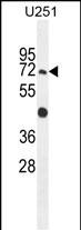
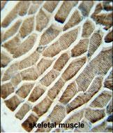
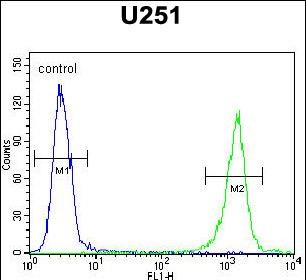
GPAA1 Antibody (N-term)
Affinity Purified Rabbit Polyclonal Antibody (Pab)
- SPECIFICATION
- CITATIONS
- PROTOCOLS
- BACKGROUND

Application
| FC, IHC-P, WB, E |
|---|---|
| Primary Accession | O43292 |
| Other Accession | NP_003792.1 |
| Reactivity | Human |
| Host | Rabbit |
| Clonality | Polyclonal |
| Isotype | Rabbit IgG |
| Calculated MW | 67623 Da |
| Antigen Region | 46-73 aa |
| Gene ID | 8733 |
|---|---|
| Other Names | Glycosylphosphatidylinositol anchor attachment 1 protein, GPI anchor attachment protein 1, GAA1 protein homolog, hGAA1, GPAA1, GAA1 |
| Target/Specificity | This GPAA1 antibody is generated from rabbits immunized with a KLH conjugated synthetic peptide between 46-73 amino acids from the N-terminal region of human GPAA1. |
| Dilution | FC~~1:10~50 IHC-P~~1:50~100 WB~~1:1000 E~~Use at an assay dependent concentration. |
| Format | Purified polyclonal antibody supplied in PBS with 0.09% (W/V) sodium azide. This antibody is purified through a protein A column, followed by peptide affinity purification. |
| Storage | Maintain refrigerated at 2-8°C for up to 2 weeks. For long term storage store at -20°C in small aliquots to prevent freeze-thaw cycles. |
| Precautions | GPAA1 Antibody (N-term) is for research use only and not for use in diagnostic or therapeutic procedures. |
| Name | GPAA1 (HGNC:4446) |
|---|---|
| Synonyms | GAA1 |
| Function | Component of the glycosylphosphatidylinositol-anchor (GPI- anchor) transamidase (GPI-T) complex that catalyzes the formation of the linkage between a proprotein and a GPI-anchor and participates in GPI anchored protein biosynthesis (PubMed:11483512, PubMed:29100095, PubMed:34576938, PubMed:35165458, PubMed:35551457, PubMed:37684232, PubMed:9468317). Binds GPI-anchor (PubMed:37684232). |
| Cellular Location | Endoplasmic reticulum membrane; Multi-pass membrane protein |
| Tissue Location | Ubiquitously expressed in fetal and adult tissues. Expressed at higher levels in fetal tissues than adult tissues |

Thousands of laboratories across the world have published research that depended on the performance of antibodies from Abcepta to advance their research. Check out links to articles that cite our products in major peer-reviewed journals, organized by research category.
info@abcepta.com, and receive a free "I Love Antibodies" mug.

Provided below are standard protocols that you may find useful for product applications.
Background
Posttranslational glycosylphosphatidylinositol (GPI) anchor attachment serves as a general mechanism for linking proteins to the cell surface membrane. The protein encoded by this gene presumably functions in GPI anchoring at the GPI transfer step. The mRNA transcript is ubiquitously expressed in both fetal and adult tissues. The anchor attachment protein 1 contains an N-terminal signal sequence, 1 cAMP- and cGMP-dependent protein kinase phosphorylation site, 1 leucine zipper pattern, 2 potential N-glycosylation sites, and 8 putative transmembrane domains.
References
Jiang, W.W., et al. Mol. Cancer 6, 74 (2007) :
Olsen, J.V., et al. Cell 127(3):635-648(2006)
Ho, J.C., et al. Int. J. Cancer 119(6):1330-1337(2006)
Vainauskas, S., et al. J. Biol. Chem. 280(16):16402-16409(2005)
Vainauskas, S., et al. J. Biol. Chem. 279(8):6540-6545(2004)
If you have used an Abcepta product and would like to share how it has performed, please click on the "Submit Review" button and provide the requested information. Our staff will examine and post your review and contact you if needed.
If you have any additional inquiries please email technical services at tech@abcepta.com.
Ordering Information
Other Products
Shipping Information
