


 Foundational characteristics of cancer include proliferation, angiogenesis, migration, evasion of apoptosis, and cellular immortality. Find key markers for these cellular processes and antibodies to detect them.
 The SUMOplot™ Analysis Program predicts and scores sumoylation sites in your protein. SUMOylation is a post-translational modification involved in various cellular processes, such as nuclear-cytosolic transport, transcriptional regulation, apoptosis, protein stability, response to stress, and progression through the cell cycle.
 The Autophagy Receptor Motif Plotter predicts and scores autophagy receptor binding sites in your protein. Identifying proteins connected to this pathway is critical to understanding the role of autophagy in physiological as well as pathological processes such as development, differentiation, neurodegenerative diseases, stress, infection, and cancer.
IBTK Antibody (Center)
Affinity Purified Rabbit Polyclonal Antibody (Pab)
- SPECIFICATION
- CITATIONS
- PROTOCOLS
- BACKGROUND

Application
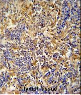
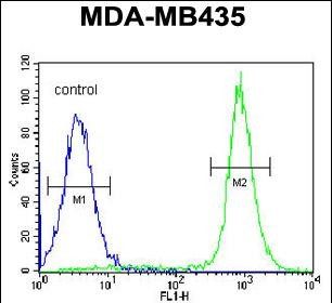
| FC, IHC-P, WB, E |
|---|---|
| Primary Accession | Q9P2D0 |
| Other Accession | NP_056340.2 |
| Reactivity | Human |
| Host | Rabbit |
| Clonality | Polyclonal |
| Isotype | Rabbit IgG |
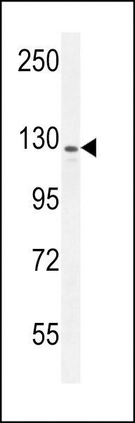
| Calculated MW | 150528 Da |
| Antigen Region | 586-613 aa |
| Gene ID | 25998 |
|---|---|
| Other Names | Inhibitor of Bruton tyrosine kinase, IBtk, IBTK, BTKI, KIAA1417 |
| Target/Specificity | This IBTK antibody is generated from rabbits immunized with a KLH conjugated synthetic peptide between 586-613 amino acids from the Central region of human IBTK. |
| Dilution | FC~~1:10~50 IHC-P~~1:50~100 WB~~1:1000 E~~Use at an assay dependent concentration. |
| Format | Purified polyclonal antibody supplied in PBS with 0.09% (W/V) sodium azide. This antibody is purified through a protein A column, followed by peptide affinity purification. |
| Storage | Maintain refrigerated at 2-8°C for up to 2 weeks. For long term storage store at -20°C in small aliquots to prevent freeze-thaw cycles. |
| Precautions | IBTK Antibody (Center) is for research use only and not for use in diagnostic or therapeutic procedures. |
| Name | IBTK |
|---|---|
| Synonyms | BTKI, KIAA1417 |
| Function | Acts as an inhibitor of BTK tyrosine kinase activity, thereby playing a role in B-cell development. Down-regulates BTK kinase activity, leading to interference with BTK-mediated calcium mobilization and NF-kappa-B-driven transcription. |
| Cellular Location | Cytoplasm. Membrane; Peripheral membrane protein. Note=Translocates to the plasma membrane upon IgM stimulation |
| Tissue Location | Expressed in DeFew, HEK293T, HeLa and in Jurkat, MC3 and NB4 lymphoid cells (at protein level). Isoform 1 is the predominant isoform expressed in all examined tissues and cell lines Highly expressed in hemopoietic tissues (fetal liver, spleen, lymph node, thymus, peripheral blood leukocytes and bone marrow). Weakly or not expressed in other tissues. |

Thousands of laboratories across the world have published research that depended on the performance of antibodies from Abcepta to advance their research. Check out links to articles that cite our products in major peer-reviewed journals, organized by research category.
info@abcepta.com, and receive a free "I Love Antibodies" mug.

Provided below are standard protocols that you may find useful for product applications.
Background
The protein encoded by this gene binds to Bruton's tyrosine kinase (BTK) and downregulates BTK's kinase activity. In addition, the encoded protein disrupts BTK-mediated calcium mobilization and negatively regulates the activation of nuclear factor-kappa-B-driven transcription.
References
Fiume, G., et al. Comput Biol Chem 33(6):434-439(2009)
Spatuzza, C., et al. Nucleic Acids Res. 36(13):4402-4416(2008)
Ewing, R.M., et al. Mol. Syst. Biol. 3, 89 (2007) :
Olsen, J.V., et al. Cell 127(3):635-648(2006)
Mungall, A.J., et al. Nature 425(6960):805-811(2003)
If you have used an Abcepta product and would like to share how it has performed, please click on the "Submit Review" button and provide the requested information. Our staff will examine and post your review and contact you if needed.
If you have any additional inquiries please email technical services at tech@abcepta.com.
Ordering Information
Other Products
Shipping Information
