


 Foundational characteristics of cancer include proliferation, angiogenesis, migration, evasion of apoptosis, and cellular immortality. Find key markers for these cellular processes and antibodies to detect them.
 The SUMOplot™ Analysis Program predicts and scores sumoylation sites in your protein. SUMOylation is a post-translational modification involved in various cellular processes, such as nuclear-cytosolic transport, transcriptional regulation, apoptosis, protein stability, response to stress, and progression through the cell cycle.
 The Autophagy Receptor Motif Plotter predicts and scores autophagy receptor binding sites in your protein. Identifying proteins connected to this pathway is critical to understanding the role of autophagy in physiological as well as pathological processes such as development, differentiation, neurodegenerative diseases, stress, infection, and cancer.
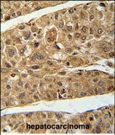
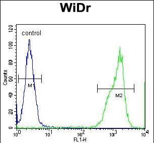
RT4I1 Antibody (C-term)
Affinity Purified Rabbit Polyclonal Antibody (Pab)

- SPECIFICATION
- CITATIONS
- PROTOCOLS
- BACKGROUND

Application
| FC, IHC-P, WB, E |
|---|---|
| Primary Accession | Q8WWV3 |
| Other Accession | NP_116119.2 |
| Reactivity | Human |
| Host | Rabbit |
| Clonality | Polyclonal |
| Isotype | Rabbit IgG |
| Calculated MW | 43590 Da |
| Antigen Region | 335-363 aa |
| Gene ID | 84816 |
|---|---|
| Other Names | Reticulon-4-interacting protein 1, mitochondrial, NOGO-interacting mitochondrial protein, RTN4IP1, NIMP |
| Target/Specificity | This RT4I1 antibody is generated from rabbits immunized with a KLH conjugated synthetic peptide between 335-363 amino acids from the C-terminal region of human RT4I1. |
| Dilution | FC~~1:10~50 IHC-P~~1:50~100 WB~~1:1000 E~~Use at an assay dependent concentration. |
| Format | Purified polyclonal antibody supplied in PBS with 0.09% (W/V) sodium azide. This antibody is purified through a protein A column, followed by peptide affinity purification. |
| Storage | Maintain refrigerated at 2-8°C for up to 2 weeks. For long term storage store at -20°C in small aliquots to prevent freeze-thaw cycles. |
| Precautions | RT4I1 Antibody (C-term) is for research use only and not for use in diagnostic or therapeutic procedures. |
| Name | RTN4IP1 {ECO:0000303|PubMed:37884807, ECO:0000312|HGNC:HGNC:18647} |
|---|---|
| Function | NAD(P)H oxidoreductase involved in the ubiquinone biosynthetic pathway (PubMed:37884807). Required for the O- methyltransferase activity of COQ3 (PubMed:37884807). Able to catalyze the oxidoreduction of 3-demethylubiquinone into 3-demethylubiquinol in vitro (PubMed:37884807). However, it is unclear if 3-demethylubiquinone constitutes a substrate in vivo (PubMed:37884807). May also play a role in the regulation of retinal ganglion cell (RGC) neurite outgrowth, and hence in the development of the inner retina and optic nerve (By similarity). Appears to be a potent inhibitor of regeneration following spinal cord injury (By similarity). |
| Cellular Location | Mitochondrion matrix. Mitochondrion outer membrane. Note=Mainly localizes to the mitochondrial matrix (PubMed:37884807). Also colocalizes with the endoplasmic reticulum HSPA5 at spots corresponding to contacts with mitochondria (PubMed:26593267) |
| Tissue Location | Widely expressed in mitochondria-enriched tissues (PubMed:12067236). Found in heart, muscle, kidney, liver, brain and placenta (PubMed:12067236). |

Thousands of laboratories across the world have published research that depended on the performance of antibodies from Abcepta to advance their research. Check out links to articles that cite our products in major peer-reviewed journals, organized by research category.
info@abcepta.com, and receive a free "I Love Antibodies" mug.

Provided below are standard protocols that you may find useful for product applications.
Background
This gene encodes a novel mitochondrial protein that interacts with reticulon 4, which is a potent inhibitor of regeneration following spinal cord injury. The interaction of reticulon 4 with mitochondrial proteins may provide insight into the mechanisms for reticulon-induced inhibition of neurite growth.
References
Rose, J. Phd, et al. Mol. Med. (2010) In press :
Mungall, A.J., et al. Nature 425(6960):805-811(2003)
Domeniconi, M., et al. Neuron 35(2):283-290(2002)
Hu, W.H., et al. J. Neurochem. 81(1):36-45(2002)
If you have used an Abcepta product and would like to share how it has performed, please click on the "Submit Review" button and provide the requested information. Our staff will examine and post your review and contact you if needed.
If you have any additional inquiries please email technical services at tech@abcepta.com.
Ordering Information
Other Products
Shipping Information
