


 Foundational characteristics of cancer include proliferation, angiogenesis, migration, evasion of apoptosis, and cellular immortality. Find key markers for these cellular processes and antibodies to detect them.
 The SUMOplot™ Analysis Program predicts and scores sumoylation sites in your protein. SUMOylation is a post-translational modification involved in various cellular processes, such as nuclear-cytosolic transport, transcriptional regulation, apoptosis, protein stability, response to stress, and progression through the cell cycle.
 The Autophagy Receptor Motif Plotter predicts and scores autophagy receptor binding sites in your protein. Identifying proteins connected to this pathway is critical to understanding the role of autophagy in physiological as well as pathological processes such as development, differentiation, neurodegenerative diseases, stress, infection, and cancer.
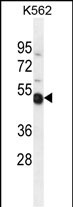
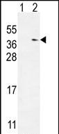
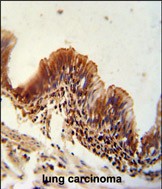
TXNDC6 Antibody (Center)
Affinity Purified Rabbit Polyclonal Antibody (Pab)
- SPECIFICATION
- CITATIONS
- PROTOCOLS
- BACKGROUND

Application
| IHC-P, WB, E |
|---|---|
| Primary Accession | Q86XW9 |
| Other Accession | NP_835231.1 |
| Reactivity | Human |
| Host | Rabbit |
| Clonality | Polyclonal |
| Isotype | Rabbit IgG |
| Calculated MW | 36856 Da |
| Antigen Region | 111-139 aa |
| Gene ID | 347736 |
|---|---|
| Other Names | Thioredoxin domain-containing protein 6, Thioredoxin-like protein 2, Txl-2, NME9, TXL2, TXNDC6 |
| Target/Specificity | This TXNDC6 antibody is generated from rabbits immunized with a KLH conjugated synthetic peptide between 111-139 amino acids from the Central region of human TXNDC6. |
| Dilution | IHC-P~~1:50~100 WB~~1:1000 E~~Use at an assay dependent concentration. |
| Format | Purified polyclonal antibody supplied in PBS with 0.09% (W/V) sodium azide. This antibody is purified through a protein A column, followed by peptide affinity purification. |
| Storage | Maintain refrigerated at 2-8°C for up to 2 weeks. For long term storage store at -20°C in small aliquots to prevent freeze-thaw cycles. |
| Precautions | TXNDC6 Antibody (Center) is for research use only and not for use in diagnostic or therapeutic procedures. |
| Name | NME9 (HGNC:21343) |
|---|---|
| Synonyms | TXL2, TXNDC6 |
| Function | May be a regulator of microtubule physiology. |
| Cellular Location | Cytoplasm, cytoskeleton, cilium axoneme {ECO:0000250|UniProtKB:A0A1L1SUL6}. Dynein axonemal particle {ECO:0000250|UniProtKB:Q6IRC5}. Note=Associated with microtubules Detected in cilia of lung epithelium, and associated with the spermatid tail and manchette. {ECO:0000250|UniProtKB:A0A1L1SUL6} |
| Tissue Location | Detected at very low levels in testis, lung and brain. |

Thousands of laboratories across the world have published research that depended on the performance of antibodies from Abcepta to advance their research. Check out links to articles that cite our products in major peer-reviewed journals, organized by research category.
info@abcepta.com, and receive a free "I Love Antibodies" mug.

Provided below are standard protocols that you may find useful for product applications.
Background
May be a regulator of microtubule physiology.
References
Lee, E.J., et al. Chest 135(2):344-352(2009)
Desvignes, T., et al. BMC Evol. Biol. 9, 256 (2009) :
Miranda-Vizuete, A., et al. Antioxid. Redox Signal. 6(1):25-40(2004)
Sadek, C.M., et al. J. Biol. Chem. 278(15):13133-13142(2003)
If you have used an Abcepta product and would like to share how it has performed, please click on the "Submit Review" button and provide the requested information. Our staff will examine and post your review and contact you if needed.
If you have any additional inquiries please email technical services at tech@abcepta.com.
Ordering Information
Shipping Information
