


 Foundational characteristics of cancer include proliferation, angiogenesis, migration, evasion of apoptosis, and cellular immortality. Find key markers for these cellular processes and antibodies to detect them.
 The SUMOplot™ Analysis Program predicts and scores sumoylation sites in your protein. SUMOylation is a post-translational modification involved in various cellular processes, such as nuclear-cytosolic transport, transcriptional regulation, apoptosis, protein stability, response to stress, and progression through the cell cycle.
 The Autophagy Receptor Motif Plotter predicts and scores autophagy receptor binding sites in your protein. Identifying proteins connected to this pathway is critical to understanding the role of autophagy in physiological as well as pathological processes such as development, differentiation, neurodegenerative diseases, stress, infection, and cancer.
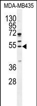
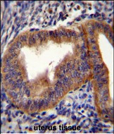
CCNB1 Antibody (Center)
Purified Rabbit Polyclonal Antibody (Pab)
- SPECIFICATION
- CITATIONS: 1
- PROTOCOLS
- BACKGROUND

Application
| WB, IHC-P, E |
|---|---|
| Primary Accession | P14635 |
| Other Accession | P30277, P24860, Q08301, Q1LZG6, NP_114172.1 |
| Reactivity | Human |
| Predicted | Bovine, Hamster, Mouse, Rat |
| Host | Rabbit |
| Clonality | Polyclonal |
| Isotype | Rabbit IgG |
| Calculated MW | 48337 Da |
| Antigen Region | 241-268 aa |
| Gene ID | 891 |
|---|---|
| Other Names | G2/mitotic-specific cyclin-B1, CCNB1, CCNB |
| Target/Specificity | This CCNB1 antibody is generated from rabbits immunized with a KLH conjugated synthetic peptide between 241-268 amino acids from the Central region of human CCNB1. |
| Dilution | WB~~1:1000 IHC-P~~1:50~100 E~~Use at an assay dependent concentration. |
| Format | Purified polyclonal antibody supplied in PBS with 0.09% (W/V) sodium azide. This antibody is prepared by Saturated Ammonium Sulfate (SAS) precipitation followed by dialysis against PBS. |
| Storage | Maintain refrigerated at 2-8°C for up to 2 weeks. For long term storage store at -20°C in small aliquots to prevent freeze-thaw cycles. |
| Precautions | CCNB1 Antibody (Center) is for research use only and not for use in diagnostic or therapeutic procedures. |
| Name | CCNB1 |
|---|---|
| Synonyms | CCNB |
| Function | Essential for the control of the cell cycle at the G2/M (mitosis) transition. |
| Cellular Location | Cytoplasm. Nucleus. Cytoplasm, cytoskeleton, microtubule organizing center, centrosome |

Provided below are standard protocols that you may find useful for product applications.
Background
The protein encoded by this gene is a regulatory protein involved in mitosis. The gene product complexes with p34(cdc2) to form the maturation-promoting factor (MPF). Two alternative transcripts have been found, a constitutively expressed transcript and a cell cycle-regulated transcript, that is expressed predominantly during G2/M phase. The different transcripts result from the use of alternate transcription initiation sites. [provided by RefSeq].
References
Kreis, N.N., et al. Oncogene 29(41):5591-5603(2010)
van Zon, W., et al. J. Cell Biol. 190(4):587-602(2010)
Harley, M.E., et al. EMBO J. 29(14):2407-2420(2010)
Olson, J.E., et al. Breast Cancer Res. Treat. (2010) In press :
Nantajit, D., et al. PLoS ONE 5 (8), E12341 (2010) :
If you have used an Abcepta product and would like to share how it has performed, please click on the "Submit Review" button and provide the requested information. Our staff will examine and post your review and contact you if needed.
If you have any additional inquiries please email technical services at tech@abcepta.com.
Ordering Information
Other Products
Shipping Information
