


 Foundational characteristics of cancer include proliferation, angiogenesis, migration, evasion of apoptosis, and cellular immortality. Find key markers for these cellular processes and antibodies to detect them.
 The SUMOplot™ Analysis Program predicts and scores sumoylation sites in your protein. SUMOylation is a post-translational modification involved in various cellular processes, such as nuclear-cytosolic transport, transcriptional regulation, apoptosis, protein stability, response to stress, and progression through the cell cycle.
 The Autophagy Receptor Motif Plotter predicts and scores autophagy receptor binding sites in your protein. Identifying proteins connected to this pathway is critical to understanding the role of autophagy in physiological as well as pathological processes such as development, differentiation, neurodegenerative diseases, stress, infection, and cancer.
KTAP2 Antibody (C-term)
Affinity Purified Rabbit Polyclonal Antibody (Pab)
- SPECIFICATION
- CITATIONS
- PROTOCOLS
- BACKGROUND

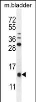
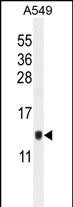
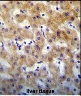
Application
| IHC-P, WB, E |
|---|---|
| Primary Accession | Q8N6L1 |
| Other Accession | B2RZC9, NP_776251.1 |
| Reactivity | Human, Mouse |
| Predicted | Rat |
| Host | Rabbit |
| Clonality | Polyclonal |
| Isotype | Rabbit IgG |
| Calculated MW | 14679 Da |
| Antigen Region | 134-162 aa |
| Gene ID | 200185 |
|---|---|
| Other Names | Keratinocyte-associated protein 2, KCP-2, KRTCAP2, KCP2 |
| Target/Specificity | This KTAP2 antibody is generated from rabbits immunized with a KLH conjugated synthetic peptide between 134-162 amino acids from the C-terminal region of human KTAP2. |
| Dilution | IHC-P~~1:10~50 WB~~1:1000 E~~Use at an assay dependent concentration. |
| Format | Purified polyclonal antibody supplied in PBS with 0.09% (W/V) sodium azide. This antibody is purified through a protein A column, followed by peptide affinity purification. |
| Storage | Maintain refrigerated at 2-8°C for up to 2 weeks. For long term storage store at -20°C in small aliquots to prevent freeze-thaw cycles. |
| Precautions | KTAP2 Antibody (C-term) is for research use only and not for use in diagnostic or therapeutic procedures. |
| Name | KRTCAP2 |
|---|---|
| Synonyms | KCP2 {ECO:0000303|PubMed:28860277} |
| Function | Subunit of STT3A-containing oligosaccharyl transferase (OST- A) complex that catalyzes the initial transfer of a defined glycan (Glc(3)Man(9)GlcNAc(2) in eukaryotes) from the lipid carrier dolichol- pyrophosphate to an asparagine residue within an Asn-X-Ser/Thr consensus motif in nascent polypeptide chains, the first step in protein N-glycosylation (PubMed:22467853, PubMed:28860277). N- glycosylation occurs cotranslationally and the complex associates with the Sec61 complex at the channel-forming translocon complex that mediates protein translocation across the endoplasmic reticulum (ER) (PubMed:22467853, PubMed:28860277). Within the OST-A complex, acts as an adapter that anchors the OST-A complex to the Sec61 complex (PubMed:28860277). May be involved in N-glycosylation of APP (amyloid- beta precursor protein) (PubMed:21768116). Can modulate gamma-secretase cleavage of APP by enhancing endoprotelysis of PSEN1 (PubMed:21768116). |
| Cellular Location | Endoplasmic reticulum. Endoplasmic reticulum membrane; Multi-pass membrane protein |
| Tissue Location | Expressed in skin, heart, placental, liver, skeletal muscle, kidney, pancreas, keratinocytes and dermal fibroblasts. |

Thousands of laboratories across the world have published research that depended on the performance of antibodies from Abcepta to advance their research. Check out links to articles that cite our products in major peer-reviewed journals, organized by research category.
info@abcepta.com, and receive a free "I Love Antibodies" mug.

Provided below are standard protocols that you may find useful for product applications.
Background
E3 ubiquitin-protein ligase which accepts ubiquitin from E2 ubiquitin-conjugating enzymes UBE2L3 and UBE2L6 in the form of a thioester and then directly transfers the ubiquitin to targeted substrates, such as UCKL1. Involved in the cytolytic activity of natural killer cells and cytotoxic T-cells.
References
Shibatani, T., et al. Biochemistry 44(16):5982-5992(2005)
Bonkobara, M., et al. Br. J. Dermatol. 148(4):654-664(2003)
If you have used an Abcepta product and would like to share how it has performed, please click on the "Submit Review" button and provide the requested information. Our staff will examine and post your review and contact you if needed.
If you have any additional inquiries please email technical services at tech@abcepta.com.
Ordering Information
Other Products
Shipping Information
