


 Foundational characteristics of cancer include proliferation, angiogenesis, migration, evasion of apoptosis, and cellular immortality. Find key markers for these cellular processes and antibodies to detect them.
 The SUMOplot™ Analysis Program predicts and scores sumoylation sites in your protein. SUMOylation is a post-translational modification involved in various cellular processes, such as nuclear-cytosolic transport, transcriptional regulation, apoptosis, protein stability, response to stress, and progression through the cell cycle.
 The Autophagy Receptor Motif Plotter predicts and scores autophagy receptor binding sites in your protein. Identifying proteins connected to this pathway is critical to understanding the role of autophagy in physiological as well as pathological processes such as development, differentiation, neurodegenerative diseases, stress, infection, and cancer.
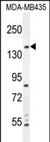
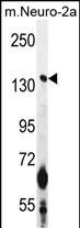
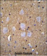
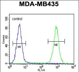
CAMSAP1 Antibody (N-term)
Affinity Purified Rabbit Polyclonal Antibody (Pab)
- SPECIFICATION
- CITATIONS
- PROTOCOLS
- BACKGROUND

Application
| FC, IHC-P, WB, E |
|---|---|
| Primary Accession | Q5T5Y3 |
| Other Accession | NP_056262.3 |
| Reactivity | Human, Mouse |
| Host | Rabbit |
| Clonality | Polyclonal |
| Isotype | Rabbit IgG |
| Calculated MW | 177972 Da |
| Antigen Region | 34-62 aa |
| Gene ID | 157922 |
|---|---|
| Other Names | Calmodulin-regulated spectrin-associated protein 1, CAMSAP1 |
| Target/Specificity | This CAMSAP1 antibody is generated from rabbits immunized with a KLH conjugated synthetic peptide between 34-62 amino acids from the N-terminal region of human CAMSAP1. |
| Dilution | FC~~1:10~50 IHC-P~~1:10~50 WB~~1:1000 E~~Use at an assay dependent concentration. |
| Format | Purified polyclonal antibody supplied in PBS with 0.09% (W/V) sodium azide. This antibody is purified through a protein A column, followed by peptide affinity purification. |
| Storage | Maintain refrigerated at 2-8°C for up to 2 weeks. For long term storage store at -20°C in small aliquots to prevent freeze-thaw cycles. |
| Precautions | CAMSAP1 Antibody (N-term) is for research use only and not for use in diagnostic or therapeutic procedures. |
| Name | CAMSAP1 |
|---|---|
| Function | Key microtubule-organizing protein that specifically binds the minus-end of non-centrosomal microtubules and regulates their dynamics and organization (PubMed:19508979, PubMed:21834987, PubMed:24117850, PubMed:24486153, PubMed:24706919). Specifically recognizes growing microtubule minus-ends and stabilizes microtubules (PubMed:24486153, PubMed:24706919). Acts on free microtubule minus-ends that are not capped by microtubule-nucleating proteins or other factors and protects microtubule minus-ends from depolymerization (PubMed:24486153, PubMed:24706919). In contrast to CAMSAP2 and CAMSAP3, tracks along the growing tips of minus-end microtubules without significantly affecting the polymerization rate: binds at the very tip of the microtubules minus-end and acts as a minus-end tracking protein (-TIP) that dissociates from microtubules after allowing tubulin incorporation (PubMed:24486153, PubMed:24706919). Through interaction with spectrin may regulate neurite outgrowth (PubMed:24117850). |
| Cellular Location | Cytoplasm, cytoskeleton. Note=Associates with the minus-end of microtubules (PubMed:24486153, PubMed:24706919). In contrast to CAMSAP2 and CAMSAP3, does not form stretches of decorated microtubule minus- ends (PubMed:24486153, PubMed:24706919). |
Research Areas
Citations (0)

Thousands of laboratories across the world have published research that depended on the performance of antibodies from Abcepta to advance their research. Check out links to articles that cite our products in major peer-reviewed journals, organized by research category.
Submit your citation using an Abcepta antibody to
info@abcepta.com, and receive a free "I Love Antibodies" mug.
info@abcepta.com, and receive a free "I Love Antibodies" mug.

Application Protocols
Provided below are standard protocols that you may find useful for product applications.
Abcepta welcomes feedback from its customers.
If you have used an Abcepta product and would like to share how it has performed, please click on the "Submit Review" button and provide the requested information. Our staff will examine and post your review and contact you if needed.
If you have any additional inquiries please email technical services at tech@abcepta.com.
$ 150.00
$ 385.00
Cat# AP11271a
Ordering Information
United States
AlbaniaAustraliaAustriaBelgiumBosnia & HerzegovinaBrazilBulgariaCanadaCentral AmericaChinaCroatiaCyprusCzech RepublicDenmarkEstoniaFinlandFranceGermanyGreeceHong KongHungaryIcelandIndiaIndonesiaIrelandIsraelItalyJapanLatviaLithuaniaLuxembourgMacedoniaMalaysiaMaltaMexicoNetherlandsNew ZealandNorwayPakistanPolandPortugalRomaniaSerbiaSingaporeSlovakiaSloveniaSouth AfricaSouth KoreaSpainSwedenSwitzerlandTaiwanTurkeyUnited KingdomUnited StatesVietnamWorldwideOthers
USA Headquarters
(888) 735-7227 / (858) 622-0099 or (858) 875-1900
Shipping Information
Domestic orders (in stock items)
Shipped out the same day. Orders placed after 1 PM (PST) will ship out the next business day.
International orders
Contact your local distributors
