


 Foundational characteristics of cancer include proliferation, angiogenesis, migration, evasion of apoptosis, and cellular immortality. Find key markers for these cellular processes and antibodies to detect them.
 The SUMOplot™ Analysis Program predicts and scores sumoylation sites in your protein. SUMOylation is a post-translational modification involved in various cellular processes, such as nuclear-cytosolic transport, transcriptional regulation, apoptosis, protein stability, response to stress, and progression through the cell cycle.
 The Autophagy Receptor Motif Plotter predicts and scores autophagy receptor binding sites in your protein. Identifying proteins connected to this pathway is critical to understanding the role of autophagy in physiological as well as pathological processes such as development, differentiation, neurodegenerative diseases, stress, infection, and cancer.
SAMD7 Antibody (Center)
Affinity Purified Rabbit Polyclonal Antibody (Pab)
- SPECIFICATION
- CITATIONS
- PROTOCOLS
- BACKGROUND

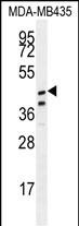
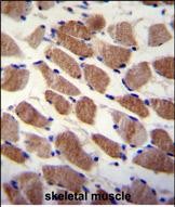
Application
| WB, IHC-P, E |
|---|---|
| Primary Accession | Q7Z3H4 |
| Other Accession | NP_872416.1 |
| Reactivity | Human |
| Host | Rabbit |
| Clonality | Polyclonal |
| Isotype | Rabbit IgG |
| Calculated MW | 49112 Da |
| Antigen Region | 202-230 aa |
| Gene ID | 344658 |
|---|---|
| Other Names | Sterile alpha motif domain-containing protein 7, SAM domain-containing protein 7, SAMD7 |
| Target/Specificity | This SAMD7 antibody is generated from rabbits immunized with a KLH conjugated synthetic peptide between 202-230 amino acids from the Central region of human SAMD7. |
| Dilution | WB~~1:1000 IHC-P~~1:10~50 E~~Use at an assay dependent concentration. |
| Format | Purified polyclonal antibody supplied in PBS with 0.09% (W/V) sodium azide. This antibody is purified through a protein A column, followed by peptide affinity purification. |
| Storage | Maintain refrigerated at 2-8°C for up to 2 weeks. For long term storage store at -20°C in small aliquots to prevent freeze-thaw cycles. |
| Precautions | SAMD7 Antibody (Center) is for research use only and not for use in diagnostic or therapeutic procedures. |
| Name | SAMD7 |
|---|---|
| Function | Component of a Polycomb group (PcG) multiprotein PRC1-like complex, essential for establishing rod photoreceptor cell identity and function by silencing nonrod gene expression in developing rod photoreceptor cells. Via its association with the PRC1-like complex, promotes epigenetic repressive marks H3K27me3 and H2AK119ub marks in nonrod genes, silencing their transcription. Represses Crx-controlled photoreceptor-specific gene expression. |
| Cellular Location | Nucleus. Cytoplasm {ECO:0000250|UniProtKB:Q8C8Y5}. Note=Co-localizes with PHC2 and RNF2 in nuclear polycomb bodies. {ECO:0000250|UniProtKB:Q8C8Y5} |
| Tissue Location | Expressed in the retina (at protein level) (PubMed:26887858). Expressed in the retinal inner and outer nuclear layers (PubMed:38272031). |
Research Areas
Citations (0)

Thousands of laboratories across the world have published research that depended on the performance of antibodies from Abcepta to advance their research. Check out links to articles that cite our products in major peer-reviewed journals, organized by research category.
Submit your citation using an Abcepta antibody to
info@abcepta.com, and receive a free "I Love Antibodies" mug.
info@abcepta.com, and receive a free "I Love Antibodies" mug.

Application Protocols
Provided below are standard protocols that you may find useful for product applications.
References
Gerhard, D.S., et al. Genome Res. 14 (10B), 2121-2127 (2004) :
Abcepta welcomes feedback from its customers.
If you have used an Abcepta product and would like to share how it has performed, please click on the "Submit Review" button and provide the requested information. Our staff will examine and post your review and contact you if needed.
If you have any additional inquiries please email technical services at tech@abcepta.com.
$ 150.00
$ 385.00
Cat# AP11411c
Ordering Information
United States
AlbaniaAustraliaAustriaBelgiumBosnia & HerzegovinaBrazilBulgariaCanadaCentral AmericaChinaCroatiaCyprusCzech RepublicDenmarkEstoniaFinlandFranceGermanyGreeceHong KongHungaryIcelandIndiaIndonesiaIrelandIsraelItalyJapanLatviaLithuaniaLuxembourgMacedoniaMalaysiaMaltaMexicoNetherlandsNew ZealandNorwayPakistanPolandPortugalRomaniaSerbiaSingaporeSlovakiaSloveniaSouth AfricaSouth KoreaSpainSwedenSwitzerlandTaiwanTurkeyUnited KingdomUnited StatesVietnamWorldwideOthers
USA Headquarters
(888) 735-7227 / (858) 622-0099 or (858) 875-1900
Shipping Information
Domestic orders (in stock items)
Shipped out the same day. Orders placed after 1 PM (PST) will ship out the next business day.
International orders
Contact your local distributors
