


 Foundational characteristics of cancer include proliferation, angiogenesis, migration, evasion of apoptosis, and cellular immortality. Find key markers for these cellular processes and antibodies to detect them.
 The SUMOplot™ Analysis Program predicts and scores sumoylation sites in your protein. SUMOylation is a post-translational modification involved in various cellular processes, such as nuclear-cytosolic transport, transcriptional regulation, apoptosis, protein stability, response to stress, and progression through the cell cycle.
 The Autophagy Receptor Motif Plotter predicts and scores autophagy receptor binding sites in your protein. Identifying proteins connected to this pathway is critical to understanding the role of autophagy in physiological as well as pathological processes such as development, differentiation, neurodegenerative diseases, stress, infection, and cancer.
SCEL Antibody (Center)
Affinity Purified Rabbit Polyclonal Antibody (Pab)

- SPECIFICATION
- CITATIONS: 1
- PROTOCOLS
- BACKGROUND

Application
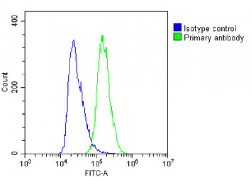
| IHC-P, WB, FC, IHC-P-Leica, E |
|---|---|
| Primary Accession | O95171 |
| Other Accession | NP_659001.2, NP_003834.3 |
| Reactivity | Human |
| Host | Rabbit |
| Clonality | Polyclonal |
| Isotype | Rabbit IgG |
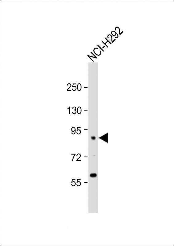
| Calculated MW | 77552 Da |
| Antigen Region | 260-289 aa |
| Gene ID | 8796 |
|---|---|
| Other Names | Sciellin, SCEL |
| Target/Specificity | This SCEL antibody is generated from rabbits immunized with a KLH conjugated synthetic peptide between 260-289 amino acids from the Central region of human SCEL. |
| Dilution | IHC-P~~N/A WB~~1:1000 FC~~1:25 IHC-P-Leica~~1:500 E~~Use at an assay dependent concentration. |
| Format | Purified polyclonal antibody supplied in PBS with 0.09% (W/V) sodium azide. This antibody is purified through a protein A column, followed by peptide affinity purification. |
| Storage | Maintain refrigerated at 2-8°C for up to 2 weeks. For long term storage store at -20°C in small aliquots to prevent freeze-thaw cycles. |
| Precautions | SCEL Antibody (Center) is for research use only and not for use in diagnostic or therapeutic procedures. |
| Name | SCEL |
|---|---|
| Function | May function in the assembly or regulation of proteins in the cornified envelope. The LIM domain may be involved in homotypic or heterotypic associations and may function to localize sciellin to the cornified envelope. |
| Cellular Location | Cytoplasm. Membrane. Note=May become cross-linked to membrane proteins by transglutaminase |
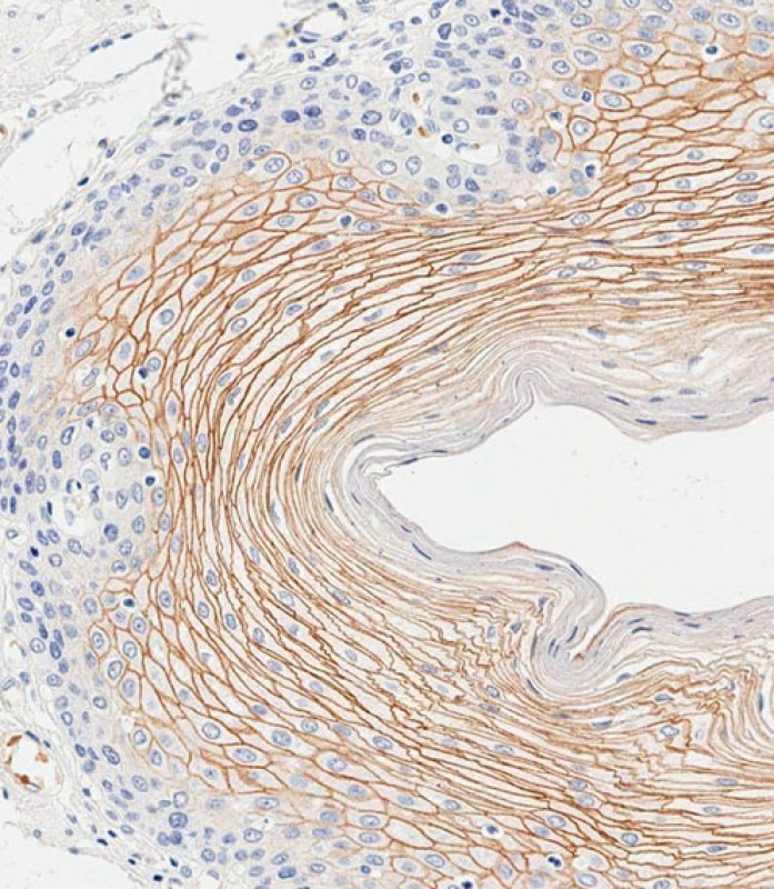
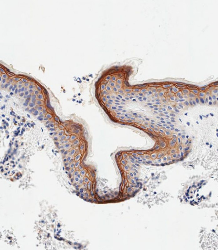
| Tissue Location | Highly expressed in esophagus. It is also expressed in keratinocytes, amniotic tissue, foreskin stratum spinosum and stratum granulosum, hair follicle and nail |

Provided below are standard protocols that you may find useful for product applications.
Background
The protein encoded by this gene is a precursor to the cornified envelope of terminally differentiated keratinocytes. This protein localizes to the periphery of cells and may function in the assembly or regulation of proteins in the cornified envelope. Transcript variants encoding different isoforms exist. A transcript variant utilizing an alternative polyA signal has been described in the literature, but its full-length nature has not been determined.
References
Pyle, A.L., et al. Cardiovasc. Pathol. 19 (2), E13-E20 (2010) :
Corona, W., et al. Anticancer Res. 24 (3A), 1417-1419 (2004) :
Champliaud, M.F., et al. J. Invest. Dermatol. 121(4):781-785(2003)
Champliaud, M.F., et al. Genomics 70(2):264-268(2000)
Champliaud, M.F., et al. J. Biol. Chem. 273(47):31547-31554(1998)
If you have used an Abcepta product and would like to share how it has performed, please click on the "Submit Review" button and provide the requested information. Our staff will examine and post your review and contact you if needed.
If you have any additional inquiries please email technical services at tech@abcepta.com.
Ordering Information
Other Products
Shipping Information
