


 Foundational characteristics of cancer include proliferation, angiogenesis, migration, evasion of apoptosis, and cellular immortality. Find key markers for these cellular processes and antibodies to detect them.
 The SUMOplot™ Analysis Program predicts and scores sumoylation sites in your protein. SUMOylation is a post-translational modification involved in various cellular processes, such as nuclear-cytosolic transport, transcriptional regulation, apoptosis, protein stability, response to stress, and progression through the cell cycle.
 The Autophagy Receptor Motif Plotter predicts and scores autophagy receptor binding sites in your protein. Identifying proteins connected to this pathway is critical to understanding the role of autophagy in physiological as well as pathological processes such as development, differentiation, neurodegenerative diseases, stress, infection, and cancer.
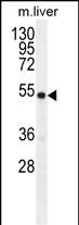
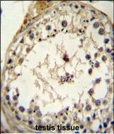
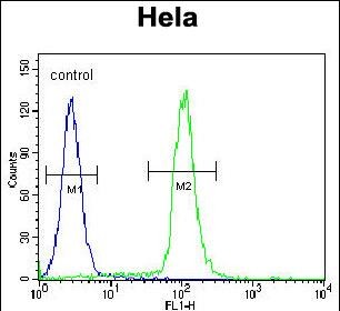
GDF6 Antibody (N-term)
Purified Rabbit Polyclonal Antibody (Pab)
- SPECIFICATION
- CITATIONS
- PROTOCOLS
- BACKGROUND

Application
| IHC-P, FC, WB, E |
|---|---|
| Primary Accession | Q6KF10 |
| Other Accession | NP_001001557.1 |
| Reactivity | Mouse |
| Host | Rabbit |
| Clonality | Polyclonal |
| Isotype | Rabbit IgG |
| Calculated MW | 50662 Da |
| Antigen Region | 31-59 aa |
| Gene ID | 392255 |
|---|---|
| Other Names | Growth/differentiation factor 6, GDF-6, Bone morphogenetic protein 13, BMP-13, Growth/differentiation factor 16, GDF6, BMP13, GDF16 |
| Target/Specificity | This GDF6 antibody is generated from rabbits immunized with a KLH conjugated synthetic peptide between 31-59 amino acids from the N-terminal region of human GDF6. |
| Dilution | IHC-P~~1:50~100 FC~~1:10~50 WB~~1:1000 E~~Use at an assay dependent concentration. |
| Format | Purified polyclonal antibody supplied in PBS with 0.09% (W/V) sodium azide. This antibody is prepared by Saturated Ammonium Sulfate (SAS) precipitation followed by dialysis against PBS. |
| Storage | Maintain refrigerated at 2-8°C for up to 2 weeks. For long term storage store at -20°C in small aliquots to prevent freeze-thaw cycles. |
| Precautions | GDF6 Antibody (N-term) is for research use only and not for use in diagnostic or therapeutic procedures. |
| Name | GDF6 |
|---|---|
| Synonyms | BMP13, GDF16 |
| Function | Growth factor that controls proliferation and cellular differentiation in the retina and bone formation. Plays a key role in regulating apoptosis during retinal development. Establishes dorsal- ventral positional information in the retina and controls the formation of the retinotectal map (PubMed:23307924). Required for normal formation of bones and joints in the limbs, skull, digits and axial skeleton. Plays a key role in establishing boundaries between skeletal elements during development. Regulation of GDF6 expression seems to be a mechanism for evolving species-specific changes in skeletal structures. Seems to positively regulate differentiation of chondrogenic tissue through the growth factor receptors subunits BMPR1A, BMPR1B, BMPR2 and ACVR2A, leading to the activation of SMAD1- SMAD5-SMAD8 complex. The regulation of chondrogenic differentiation is inhibited by NOG (PubMed:26643732). Also involved in the induction of adipogenesis from mesenchymal stem cells. This mechanism acts through the growth factor receptors subunits BMPR1A, BMPR2 and ACVR2A and the activation of SMAD1-SMAD5-SMAD8 complex and MAPK14/p38 (By similarity). |
| Cellular Location | Secreted. |

Thousands of laboratories across the world have published research that depended on the performance of antibodies from Abcepta to advance their research. Check out links to articles that cite our products in major peer-reviewed journals, organized by research category.
info@abcepta.com, and receive a free "I Love Antibodies" mug.

Provided below are standard protocols that you may find useful for product applications.
Background
This gene encodes a member of the bone morphogenetic protein (BMP) family and the TGF-beta superfamily of secreted signaling molecules. It is required for normal formation of some bones and joints in the limbs, skull, and axial skeleton. Mutations in this gene result in colobomata, which are congenital abnormalities in ocular development, and in Klippel-Feil syndrome (KFS), which is a congenital disorder of spinal segmentation.
References
Gonzalez-Rodriguez, J., et al. Br J Ophthalmol 94(8):1100-1104(2010)
Mikic, B., et al. J. Orthop. Res. 27(12):1603-1611(2009)
Asai-Coakwell, M., et al. Hum. Mol. Genet. 18(6):1110-1121(2009)
Zhang, X., et al. Mol. Vis. 15, 2911-2918 (2009) :
Shen, B., et al. Int. J. Biol. Sci. 5(2):192-200(2009)
If you have used an Abcepta product and would like to share how it has performed, please click on the "Submit Review" button and provide the requested information. Our staff will examine and post your review and contact you if needed.
If you have any additional inquiries please email technical services at tech@abcepta.com.
Ordering Information
Other Products
Shipping Information
