


 Foundational characteristics of cancer include proliferation, angiogenesis, migration, evasion of apoptosis, and cellular immortality. Find key markers for these cellular processes and antibodies to detect them.
 The SUMOplot™ Analysis Program predicts and scores sumoylation sites in your protein. SUMOylation is a post-translational modification involved in various cellular processes, such as nuclear-cytosolic transport, transcriptional regulation, apoptosis, protein stability, response to stress, and progression through the cell cycle.
 The Autophagy Receptor Motif Plotter predicts and scores autophagy receptor binding sites in your protein. Identifying proteins connected to this pathway is critical to understanding the role of autophagy in physiological as well as pathological processes such as development, differentiation, neurodegenerative diseases, stress, infection, and cancer.
NIX Antibody (Center)
Affinity Purified Rabbit Polyclonal Antibody (Pab)
- SPECIFICATION
- CITATIONS
- PROTOCOLS
- BACKGROUND

Application
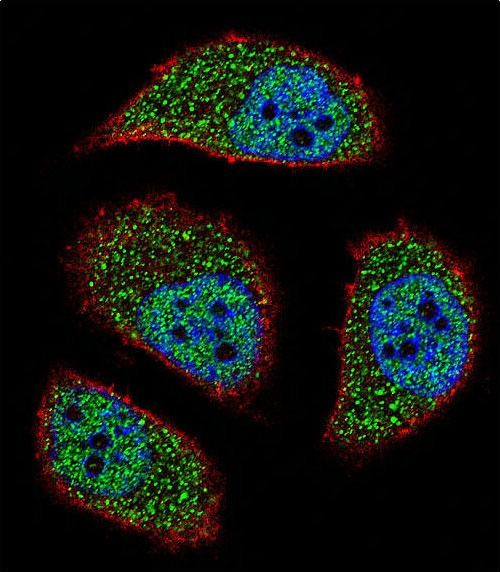
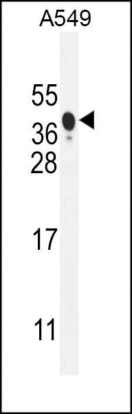
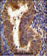
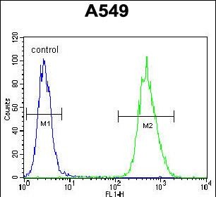
| FC, IF, IHC-P, WB, E |
|---|---|
| Primary Accession | O60238 |
| Other Accession | Q9Z2F7, Q3T013, NP_004322.1 |
| Reactivity | Human |
| Predicted | Bovine, Mouse |
| Host | Rabbit |
| Clonality | Polyclonal |
| Isotype | Rabbit IgG |
| Calculated MW | 23930 Da |
| Antigen Region | 52-81 aa |
| Gene ID | 665 |
|---|---|
| Other Names | BCL2/adenovirus E1B 19 kDa protein-interacting protein 3-like, Adenovirus E1B19K-binding protein B5, BCL2/adenovirus E1B 19 kDa protein-interacting protein 3A, NIP3-like protein X, NIP3L, BNIP3L, BNIP3A, BNIP3H, NIX |
| Target/Specificity | This NIX antibody is generated from rabbits immunized with a KLH conjugated synthetic peptide between 52-81 amino acids from the Central region of human NIX. |
| Dilution | FC~~1:10~50 IF~~1:10~50 IHC-P~~1:50~100 WB~~1:1000 E~~Use at an assay dependent concentration. |
| Format | Purified polyclonal antibody supplied in PBS with 0.09% (W/V) sodium azide. This antibody is purified through a protein A column, followed by peptide affinity purification. |
| Storage | Maintain refrigerated at 2-8°C for up to 2 weeks. For long term storage store at -20°C in small aliquots to prevent freeze-thaw cycles. |
| Precautions | NIX Antibody (Center) is for research use only and not for use in diagnostic or therapeutic procedures. |
| Name | BNIP3L |
|---|---|
| Synonyms | BNIP3A, BNIP3H, NIX |
| Function | Induces apoptosis. Interacts with viral and cellular anti- apoptosis proteins. Can overcome the suppressors BCL-2 and BCL-XL, although high levels of BCL-XL expression will inhibit apoptosis. Inhibits apoptosis induced by BNIP3. Involved in mitochondrial quality control via its interaction with SPATA18/MIEAP: in response to mitochondrial damage, participates in mitochondrial protein catabolic process (also named MALM) leading to the degradation of damaged proteins inside mitochondria. The physical interaction of SPATA18/MIEAP, BNIP3 and BNIP3L/NIX at the mitochondrial outer membrane regulates the opening of a pore in the mitochondrial double membrane in order to mediate the translocation of lysosomal proteins from the cytoplasm to the mitochondrial matrix. May function as a tumor suppressor. |
| Cellular Location | Nucleus envelope. Endoplasmic reticulum. Mitochondrion outer membrane. Membrane; Single-pass membrane protein. Note=Colocalizes with SPATA18 at the mitochondrion outer membrane |

Thousands of laboratories across the world have published research that depended on the performance of antibodies from Abcepta to advance their research. Check out links to articles that cite our products in major peer-reviewed journals, organized by research category.
info@abcepta.com, and receive a free "I Love Antibodies" mug.

Provided below are standard protocols that you may find useful for product applications.
Background
This gene is a member of the BCL2/adenovirus E1B 19 kd-interacting protein (BNIP) family. It interacts with the E1B 19 kDa protein which is responsible for the protection of virally-induced cell death, as well as E1B 19 kDa-like sequences of BCL2, also an apoptotic protector. The protein encoded by this gene is a functional homolog of BNIP3, a proapoptotic protein. This protein may function simultaneously with BNIP3 and may play a role in tumor suppression.
References
Novak, I., et al. EMBO Rep. 11(1):45-51(2010)
Bellot, G., et al. Mol. Cell. Biol. 29(10):2570-2581(2009)
Wang, L., et al. Cancer Epidemiol. Biomarkers Prev. 17(12):3558-3566(2008)
Papandreou, I., et al. Cell Death Differ. 15(10):1572-1581(2008)
Liu, W., et al. Neoplasia 10(8):897-907(2008)
If you have used an Abcepta product and would like to share how it has performed, please click on the "Submit Review" button and provide the requested information. Our staff will examine and post your review and contact you if needed.
If you have any additional inquiries please email technical services at tech@abcepta.com.

Ordering Information
Other Products
Shipping Information
