


 Foundational characteristics of cancer include proliferation, angiogenesis, migration, evasion of apoptosis, and cellular immortality. Find key markers for these cellular processes and antibodies to detect them.
 The SUMOplot™ Analysis Program predicts and scores sumoylation sites in your protein. SUMOylation is a post-translational modification involved in various cellular processes, such as nuclear-cytosolic transport, transcriptional regulation, apoptosis, protein stability, response to stress, and progression through the cell cycle.
 The Autophagy Receptor Motif Plotter predicts and scores autophagy receptor binding sites in your protein. Identifying proteins connected to this pathway is critical to understanding the role of autophagy in physiological as well as pathological processes such as development, differentiation, neurodegenerative diseases, stress, infection, and cancer.
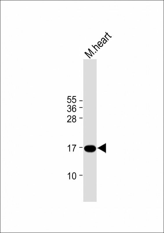
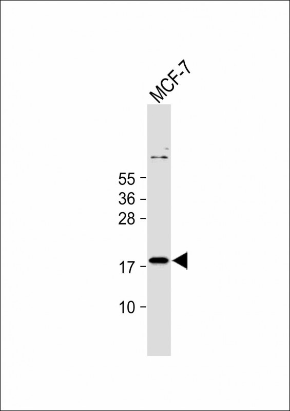
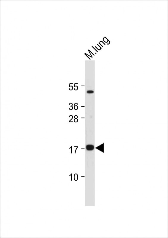
IER3 Antibody (N-term)
Affinity Purified Rabbit Polyclonal Antibody (Pab)
- SPECIFICATION
- CITATIONS: 1
- PROTOCOLS
- BACKGROUND

Application
| WB, E |
|---|---|
| Primary Accession | P46695 |
| Other Accession | NP_003888.2 |
| Reactivity | Human, Mouse |
| Host | Rabbit |
| Clonality | Polyclonal |
| Isotype | Rabbit IgG |
| Calculated MW | 16903 Da |
| Antigen Region | 38-65 aa |
| Gene ID | 8870 |
|---|---|
| Other Names | Radiation-inducible immediate-early gene IEX-1, Differentiation-dependent gene 2 protein, Protein DIF-2, Immediate early protein GLY96, Immediate early response 3 protein, PACAP-responsive gene 1 protein, Protein PRG1, IER3, DIF2, IEX1, PRG1 |
| Target/Specificity | This IER3 antibody is generated from rabbits immunized with a KLH conjugated synthetic peptide between 38-65 amino acids from the N-terminal region of human IER3. |
| Dilution | WB~~1:2000 E~~Use at an assay dependent concentration. |
| Format | Purified polyclonal antibody supplied in PBS with 0.09% (W/V) sodium azide. This antibody is purified through a protein A column, followed by peptide affinity purification. |
| Storage | Maintain refrigerated at 2-8°C for up to 2 weeks. For long term storage store at -20°C in small aliquots to prevent freeze-thaw cycles. |
| Precautions | IER3 Antibody (N-term) is for research use only and not for use in diagnostic or therapeutic procedures. |
| Name | IER3 |
|---|---|
| Synonyms | DIF2, IEX1, PRG1 |
| Function | May play a role in the ERK signaling pathway by inhibiting the dephosphorylation of ERK by phosphatase PP2A-PPP2R5C holoenzyme. Also acts as an ERK downstream effector mediating survival. As a member of the NUPR1/RELB/IER3 survival pathway, may provide pancreatic ductal adenocarcinoma with remarkable resistance to cell stress, such as starvation or gemcitabine treatment. |
| Cellular Location | Membrane; Single- pass type II membrane protein |

Provided below are standard protocols that you may find useful for product applications.
Background
This gene functions in the protection of cells from Fas- or tumor necrosis factor type alpha-induced apoptosis. Partially degraded and unspliced transcripts are found after virus infection in vitro, but these transcripts are not found in vivo and do not generate a valid protein.
References
Shahid, M., et al. Hypertension 56(4):705-712(2010)
Ucisik-Akkaya, E., et al. Mol. Hum. Reprod. 16(10):770-777(2010)
Fellay, J., et al. PLoS Genet. 5 (12), E1000791 (2009) :
Do, T.N., et al. Cancer Genet. Cytogenet. 195(1):31-36(2009)
Barcellos, L.F., et al. PLoS Genet. 5 (10), E1000696 (2009) :
If you have used an Abcepta product and would like to share how it has performed, please click on the "Submit Review" button and provide the requested information. Our staff will examine and post your review and contact you if needed.
If you have any additional inquiries please email technical services at tech@abcepta.com.
Ordering Information
Other Products
Shipping Information
