


 Foundational characteristics of cancer include proliferation, angiogenesis, migration, evasion of apoptosis, and cellular immortality. Find key markers for these cellular processes and antibodies to detect them.
 The SUMOplot™ Analysis Program predicts and scores sumoylation sites in your protein. SUMOylation is a post-translational modification involved in various cellular processes, such as nuclear-cytosolic transport, transcriptional regulation, apoptosis, protein stability, response to stress, and progression through the cell cycle.
 The Autophagy Receptor Motif Plotter predicts and scores autophagy receptor binding sites in your protein. Identifying proteins connected to this pathway is critical to understanding the role of autophagy in physiological as well as pathological processes such as development, differentiation, neurodegenerative diseases, stress, infection, and cancer.
PTPN20A Antibody (Center)
Affinity Purified Rabbit Polyclonal Antibody (Pab)

- SPECIFICATION
- CITATIONS
- PROTOCOLS
- BACKGROUND

Application
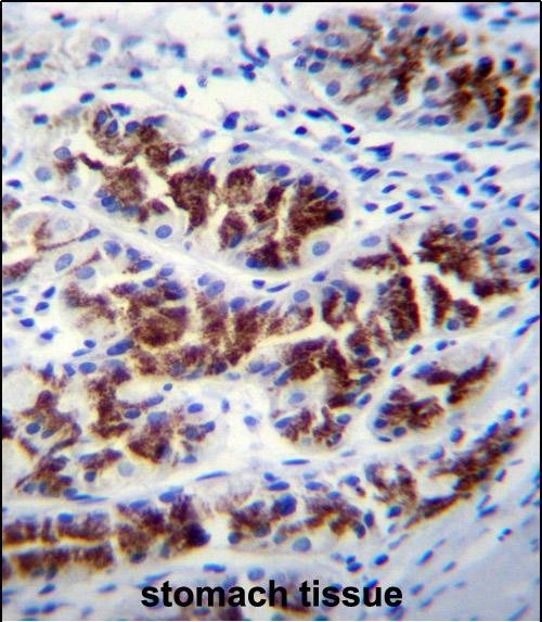
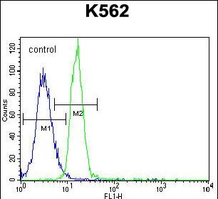
| FC, IHC-P, WB, E |
|---|---|
| Primary Accession | Q4JDL3 |
| Other Accession | NP_001035817.1, NP_001035853.1, NP_001035849.1 |
| Reactivity | Human |
| Host | Rabbit |
| Clonality | Polyclonal |
| Isotype | Rabbit IgG |
| Calculated MW | 48423 Da |
| Antigen Region | 178-207 aa |
| Gene ID | 26095 |
|---|---|
| Other Names | Tyrosine-protein phosphatase non-receptor type 20, hPTPN20, PTPN20A |
| Target/Specificity | This PTPN20A antibody is generated from rabbits immunized with a KLH conjugated synthetic peptide between 178-207 amino acids from the Central region of human PTPN20A. |
| Dilution | FC~~1:10~50 IHC-P~~1:10~50 WB~~1:1000 E~~Use at an assay dependent concentration. |
| Format | Purified polyclonal antibody supplied in PBS with 0.09% (W/V) sodium azide. This antibody is purified through a protein A column, followed by peptide affinity purification. |
| Storage | Maintain refrigerated at 2-8°C for up to 2 weeks. For long term storage store at -20°C in small aliquots to prevent freeze-thaw cycles. |
| Precautions | PTPN20A Antibody (Center) is for research use only and not for use in diagnostic or therapeutic procedures. |
| Name | PTPN20 (HGNC:23423) |
|---|---|
| Function | Tyrosine-protein phosphatase targeted to sites of actin polymerization in response of varied extracellular stimuli. Has tyrosine phosphatase activity towards various tyrosyl phosphorylated substrates. |
| Cellular Location | Nucleus. Cytoplasm. Cytoplasm, cytoskeleton, microtubule organizing center, centrosome Note=Colocalizes with the microtubule-organizing center and intracellular membrane compartments |
| Tissue Location | Present in many cell lines (at protein level). Widely expressed. |

Thousands of laboratories across the world have published research that depended on the performance of antibodies from Abcepta to advance their research. Check out links to articles that cite our products in major peer-reviewed journals, organized by research category.
info@abcepta.com, and receive a free "I Love Antibodies" mug.

Provided below are standard protocols that you may find useful for product applications.
Background
The product of this gene belongs to the family of classical tyrosine-specific protein tyrosine phosphatases. Many protein tyrosine phosphatases have been shown to regulate fundamental cellular processes and several are mutated in human diseases. Chromosome 10q contains a segmental duplication resulting in multiple copies of the protein tyrosine phosphatase, non-receptor type 20 gene. The two nearly identical copies are designated as PTPN20A and PTPN20B. A third copy is only partially duplicated and contains a pseudogene, designated as PTPN20C. This gene encodes the more telomeric copy, PTPN20B. Multiple alternatively spliced transcript variants encoding different isoforms have been identified.
References
Matsuoka, S., et al. Science 316(5828):1160-1166(2007)
Fodero-Tavoletti, M.T., et al. Biochem. J. 389 (PT 2), 343-354 (2005) :
If you have used an Abcepta product and would like to share how it has performed, please click on the "Submit Review" button and provide the requested information. Our staff will examine and post your review and contact you if needed.
If you have any additional inquiries please email technical services at tech@abcepta.com.
Ordering Information
Shipping Information
