


 Foundational characteristics of cancer include proliferation, angiogenesis, migration, evasion of apoptosis, and cellular immortality. Find key markers for these cellular processes and antibodies to detect them.
 The SUMOplot™ Analysis Program predicts and scores sumoylation sites in your protein. SUMOylation is a post-translational modification involved in various cellular processes, such as nuclear-cytosolic transport, transcriptional regulation, apoptosis, protein stability, response to stress, and progression through the cell cycle.
 The Autophagy Receptor Motif Plotter predicts and scores autophagy receptor binding sites in your protein. Identifying proteins connected to this pathway is critical to understanding the role of autophagy in physiological as well as pathological processes such as development, differentiation, neurodegenerative diseases, stress, infection, and cancer.
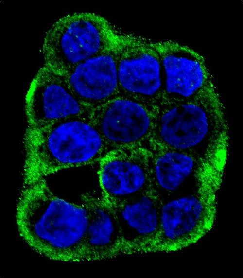
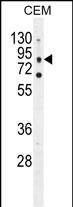
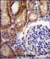
CLCN7 Antibody (C-term)
Affinity Purified Rabbit Polyclonal Antibody (Pab)
- SPECIFICATION
- CITATIONS: 1
- PROTOCOLS
- BACKGROUND

Application
| WB, IF, IHC-P, E |
|---|---|
| Primary Accession | P51798 |
| Other Accession | P51799, O70496, Q4PKH3, NP_001278.1 |
| Reactivity | Human |
| Predicted | Bovine, Mouse, Rat |
| Host | Rabbit |
| Clonality | Polyclonal |
| Isotype | Rabbit IgG |
| Calculated MW | 88679 Da |
| Antigen Region | 692-720 aa |
| Gene ID | 1186 |
|---|---|
| Other Names | H(+)/Cl(-) exchange transporter 7, Chloride channel 7 alpha subunit, Chloride channel protein 7, ClC-7, CLCN7 |
| Target/Specificity | This CLCN7 antibody is generated from rabbits immunized with a KLH conjugated synthetic peptide between 692-720 amino acids from the C-terminal region of human CLCN7. |
| Dilution | WB~~1:1000 IF~~1:10~50 IHC-P~~1:10~50 E~~Use at an assay dependent concentration. |
| Format | Purified polyclonal antibody supplied in PBS with 0.09% (W/V) sodium azide. This antibody is purified through a protein A column, followed by peptide affinity purification. |
| Storage | Maintain refrigerated at 2-8°C for up to 2 weeks. For long term storage store at -20°C in small aliquots to prevent freeze-thaw cycles. |
| Precautions | CLCN7 Antibody (C-term) is for research use only and not for use in diagnostic or therapeutic procedures. |
| Name | CLCN7 (HGNC:2025) |
|---|---|
| Function | Slowly voltage-gated channel mediating the exchange of chloride ions against protons (PubMed:18449189, PubMed:21527911). Functions as antiporter and contributes to the acidification of the lysosome lumen and may be involved in maintaining lysosomal pH (PubMed:18449189, PubMed:21527911, PubMed:31155284). The CLC channel family contains both chloride channels and proton-coupled anion transporters that exchange chloride or another anion for protons (By similarity). The presence of conserved gating glutamate residues is typical for family members that function as antiporters (By similarity). |
| Cellular Location | Lysosome membrane; Multi-pass membrane protein |
| Tissue Location | Brain and kidney.. |

Provided below are standard protocols that you may find useful for product applications.
Background
The product of this gene belongs to the CLC chloride channel family of proteins. Chloride channels play important roles in the plasma membrane and in intracellular organelles. This gene encodes chloride channel 7. Defects in this gene are the cause of osteopetrosis autosomal recessive type 4 (OPTB4), also called infantile malignant osteopetrosis type 2 as well as the cause of autosomal dominant osteopetrosis type 2 (OPTA2), also called autosomal dominant Albers-Schonberg disease or marble disease autosoml dominant. Osteopetrosis is a rare genetic disease characterized by abnormally dense bone, due to defective resorption of immature bone. OPTA2 is the most common form of osteopetrosis, occurring in adolescence or adulthood.
References
Furthner, D., et al. Klin Padiatr 222(3):180-183(2010)
Phadke, S.R., et al. Indian J. Med. Res. 131, 508-514 (2010) :
Pangrazio, A., et al. Hum. Mutat. 31 (1), E1071-E1080 (2010) :
Kajiya, H., et al. Pflugers Arch. 458(6):1049-1059(2009)
Mazzolari, E., et al. Am. J. Hematol. 84(8):473-479(2009)
If you have used an Abcepta product and would like to share how it has performed, please click on the "Submit Review" button and provide the requested information. Our staff will examine and post your review and contact you if needed.
If you have any additional inquiries please email technical services at tech@abcepta.com.
Ordering Information
Other Products
Shipping Information
