


 Foundational characteristics of cancer include proliferation, angiogenesis, migration, evasion of apoptosis, and cellular immortality. Find key markers for these cellular processes and antibodies to detect them.
 The SUMOplot™ Analysis Program predicts and scores sumoylation sites in your protein. SUMOylation is a post-translational modification involved in various cellular processes, such as nuclear-cytosolic transport, transcriptional regulation, apoptosis, protein stability, response to stress, and progression through the cell cycle.
 The Autophagy Receptor Motif Plotter predicts and scores autophagy receptor binding sites in your protein. Identifying proteins connected to this pathway is critical to understanding the role of autophagy in physiological as well as pathological processes such as development, differentiation, neurodegenerative diseases, stress, infection, and cancer.
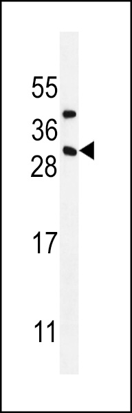
CD8B1 Antibody (C-term)
Affinity Purified Rabbit Polyclonal Antibody (Pab)
- SPECIFICATION
- CITATIONS
- PROTOCOLS
- BACKGROUND

Application
| WB, E |
|---|---|
| Primary Accession | P10966 |
| Other Accession | A6NJW9, NP_004922 |
| Reactivity | Human |
| Host | Rabbit |
| Clonality | Polyclonal |
| Isotype | Rabbit IgG |
| Calculated MW | 23722 Da |
| Antigen Region | 141-167 aa |
| Gene ID | 926 |
|---|---|
| Other Names | T-cell surface glycoprotein CD8 beta chain, CD8b, CD8B, CD8B1 |
| Target/Specificity | This CD8B1 antibody is generated from rabbits immunized with a KLH conjugated synthetic peptide between 141-167 amino acids from the C-terminal region of human CD8B1. |
| Dilution | WB~~1:1000 E~~Use at an assay dependent concentration. |
| Format | Purified polyclonal antibody supplied in PBS with 0.09% (W/V) sodium azide. This antibody is purified through a protein A column, followed by peptide affinity purification. |
| Storage | Maintain refrigerated at 2-8°C for up to 2 weeks. For long term storage store at -20°C in small aliquots to prevent freeze-thaw cycles. |
| Precautions | CD8B1 Antibody (C-term) is for research use only and not for use in diagnostic or therapeutic procedures. |
| Name | CD8B |
|---|---|
| Synonyms | CD8B1 |
| Function | Integral membrane glycoprotein that plays an essential role in the immune response and serves multiple functions in responses against both external and internal offenses. In T-cells, functions primarily as a coreceptor for MHC class I molecule:peptide complex. The antigens presented by class I peptides are derived from cytosolic proteins while class II derived from extracellular proteins. Interacts simultaneously with the T-cell receptor (TCR) and the MHC class I proteins presented by antigen presenting cells (APCs). In turn, recruits the Src kinase LCK to the vicinity of the TCR-CD3 complex. A palmitoylation site in the cytoplasmic tail of CD8B chain contributes to partitioning of CD8 into the plasma membrane lipid rafts where signaling proteins are enriched. Once LCK recruited, it initiates different intracellular signaling pathways by phosphorylating various substrates ultimately leading to lymphokine production, motility, adhesion and activation of cytotoxic T-lymphocytes (CTLs). Additionally, plays a critical role in thymic selection of CD8+ T- cells. |
| Cellular Location | [Isoform 1]: Cell membrane; Single-pass type I membrane protein. Note=Requires the partner CD8A for efficient cell surface expression (PubMed:3145196). The heterodimer CD8A/CD8B localizes to lipid rafts due to CD8B cytoplasmic tail palmitoylation. [Isoform 3]: Secreted. [Isoform 5]: Cell membrane; Single- pass type I membrane protein [Isoform 7]: Secreted. |
| Tissue Location | Isoform 1, isoform 3, isoform 5, isoform 6, isoform 7 and isoform 8 are expressed in both thymus and peripheral CD8+ T- cells. Expression of isoform 1 is higher in thymus CD8+ T-cells than in peripheral CD8+ T-cells. Expression of isoform 6 is higher in peripheral CD8+ T-cells than in thymus CD8+ T-cells |

Thousands of laboratories across the world have published research that depended on the performance of antibodies from Abcepta to advance their research. Check out links to articles that cite our products in major peer-reviewed journals, organized by research category.
info@abcepta.com, and receive a free "I Love Antibodies" mug.

Provided below are standard protocols that you may find useful for product applications.
Background
The CD8 antigen is a cell surface glycoprotein found on most cytotoxic T lymphocytes that mediates efficient cell-cell interactions within the immune system. The CD8 antigen, acting as a coreceptor, and the T-cell receptor on the T lymphocyte recognize antigens displayed by an antigen presenting cell (APC) in the context of class I MHC molecules. The functional coreceptor is either a homodimer composed of two alpha chains, or a heterodimer composed of one alpha and one beta chain. Both alpha and beta chains share significant homology to immunoglobulin variable light chains. This gene encodes the CD8 beta chain isoforms. Multiple alternatively spliced transcript variants encoding distinct membrane associated or secreted isoforms have been described. A pseudogene, also located on chromosome 2, has been identified.
References
Rose, J.E., et al. Mol. Med. 16 (7-8), 247-253 (2010) :
Chen, M.L., et al. Eur. J. Immunol. 39(12):3423-3435(2009)
Thakral, D., et al. J. Immunol. 180(11):7431-7442(2008)
Stove, V., et al. J. Virol. 79(17):11422-11433(2005)
Moody, A.M., et al. J. Biol. Chem. 278(9):7240-7246(2003)
If you have used an Abcepta product and would like to share how it has performed, please click on the "Submit Review" button and provide the requested information. Our staff will examine and post your review and contact you if needed.
If you have any additional inquiries please email technical services at tech@abcepta.com.
Ordering Information
Other Products
Shipping Information
