


 Foundational characteristics of cancer include proliferation, angiogenesis, migration, evasion of apoptosis, and cellular immortality. Find key markers for these cellular processes and antibodies to detect them.
 The SUMOplot™ Analysis Program predicts and scores sumoylation sites in your protein. SUMOylation is a post-translational modification involved in various cellular processes, such as nuclear-cytosolic transport, transcriptional regulation, apoptosis, protein stability, response to stress, and progression through the cell cycle.
 The Autophagy Receptor Motif Plotter predicts and scores autophagy receptor binding sites in your protein. Identifying proteins connected to this pathway is critical to understanding the role of autophagy in physiological as well as pathological processes such as development, differentiation, neurodegenerative diseases, stress, infection, and cancer.
PHF20 Antibody (N-term)
Affinity Purified Rabbit Polyclonal Antibody (Pab)
- SPECIFICATION
- CITATIONS
- PROTOCOLS
- BACKGROUND

Application
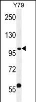
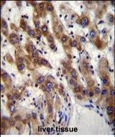
| IHC-P, WB, E |
|---|---|
| Primary Accession | Q9BVI0 |
| Other Accession | Q8BLG0, NP_057520.2 |
| Reactivity | Human |
| Predicted | Mouse |
| Host | Rabbit |
| Clonality | Polyclonal |
| Isotype | Rabbit IgG |
| Calculated MW | 115386 Da |
| Antigen Region | 123-151 aa |
| Gene ID | 51230 |
|---|---|
| Other Names | PHD finger protein 20, Glioma-expressed antigen 2, Hepatocellular carcinoma-associated antigen 58, Novel zinc finger protein, Transcription factor TZP, PHF20, C20orf104, GLEA2, HCA58, NZF, TZP |
| Target/Specificity | This PHF20 antibody is generated from rabbits immunized with a KLH conjugated synthetic peptide between 123-151 amino acids from the N-terminal region of human PHF20. |
| Dilution | IHC-P~~1:10~50 WB~~1:1000 E~~Use at an assay dependent concentration. |
| Format | Purified polyclonal antibody supplied in PBS with 0.09% (W/V) sodium azide. This antibody is purified through a protein A column, followed by peptide affinity purification. |
| Storage | Maintain refrigerated at 2-8°C for up to 2 weeks. For long term storage store at -20°C in small aliquots to prevent freeze-thaw cycles. |
| Precautions | PHF20 Antibody (N-term) is for research use only and not for use in diagnostic or therapeutic procedures. |
| Name | PHF20 |
|---|---|
| Synonyms | C20orf104, GLEA2, HCA58, NZF, TZP |
| Function | Methyllysine-binding protein, component of the MOF histone acetyltransferase protein complex. Not required for maintaining the global histone H4 'Lys-16' acetylation (H4K16ac) levels or locus specific histone acetylation, but instead works downstream in transcriptional regulation of MOF target genes (By similarity). As part of the NSL complex it may be involved in acetylation of nucleosomal histone H4 on several lysine residues. Contributes to methyllysine- dependent p53/TP53 stabilization and up-regulation after DNA damage. |
| Cellular Location | Nucleus. |
| Tissue Location | Expressed in heart, kidney, liver, lung, pancreas, placenta, spleen and testis. Not expressed in brain, skeletal muscle, colon, ovary, prostate, small intestine and thymus. Expressed in colon and ovary cancer cell lines while it is not expressed in the respective normal tissues. |

Thousands of laboratories across the world have published research that depended on the performance of antibodies from Abcepta to advance their research. Check out links to articles that cite our products in major peer-reviewed journals, organized by research category.
info@abcepta.com, and receive a free "I Love Antibodies" mug.

Provided below are standard protocols that you may find useful for product applications.
Background
PHF20 is possible a transcription factor.
References
Bankovic, J., et al. Lung Cancer 67(2):151-159(2010)
Heisel, S.M., et al. PLoS ONE 3 (5), E2164 (2008) :
Olsen, J.V., et al. Cell 127(3):635-648(2006)
Pallasch, C.P., et al. Int. J. Cancer 117(3):456-459(2005)
Dou, Y., et al. Cell 121(6):873-885(2005)
If you have used an Abcepta product and would like to share how it has performed, please click on the "Submit Review" button and provide the requested information. Our staff will examine and post your review and contact you if needed.
If you have any additional inquiries please email technical services at tech@abcepta.com.
Ordering Information
Other Products
Shipping Information
