


 Foundational characteristics of cancer include proliferation, angiogenesis, migration, evasion of apoptosis, and cellular immortality. Find key markers for these cellular processes and antibodies to detect them.
 The SUMOplot™ Analysis Program predicts and scores sumoylation sites in your protein. SUMOylation is a post-translational modification involved in various cellular processes, such as nuclear-cytosolic transport, transcriptional regulation, apoptosis, protein stability, response to stress, and progression through the cell cycle.
 The Autophagy Receptor Motif Plotter predicts and scores autophagy receptor binding sites in your protein. Identifying proteins connected to this pathway is critical to understanding the role of autophagy in physiological as well as pathological processes such as development, differentiation, neurodegenerative diseases, stress, infection, and cancer.
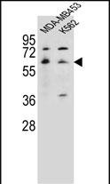
HTR3A Antibody (N-term)
Affinity Purified Rabbit Polyclonal Antibody (Pab)
- SPECIFICATION
- CITATIONS
- PROTOCOLS
- BACKGROUND

Application
| WB, E |
|---|---|
| Primary Accession | P46098 |
| Other Accession | NP_998786.2, NP_000860.2, NP_001155244.1 |
| Reactivity | Human |
| Host | Rabbit |
| Clonality | Polyclonal |
| Isotype | Rabbit IgG |
| Calculated MW | 55280 Da |
| Antigen Region | 30-58 aa |
| Gene ID | 3359 |
|---|---|
| Other Names | 5-hydroxytryptamine receptor 3A, 5-HT3-A, 5-HT3A, 5-hydroxytryptamine receptor 3, 5-HT-3, 5-HT3R, Serotonin receptor 3A, Serotonin-gated ion channel receptor, HTR3A, 5HT3R, HTR3 |
| Target/Specificity | This HTR3A antibody is generated from rabbits immunized with a KLH conjugated synthetic peptide between 30-58 amino acids from the N-terminal region of human HTR3A. |
| Dilution | WB~~1:1000 E~~Use at an assay dependent concentration. |
| Format | Purified polyclonal antibody supplied in PBS with 0.09% (W/V) sodium azide. This antibody is purified through a protein A column, followed by peptide affinity purification. |
| Storage | Maintain refrigerated at 2-8°C for up to 2 weeks. For long term storage store at -20°C in small aliquots to prevent freeze-thaw cycles. |
| Precautions | HTR3A Antibody (N-term) is for research use only and not for use in diagnostic or therapeutic procedures. |
| Name | HTR3A (HGNC:5297) |
|---|---|
| Synonyms | 5HT3R, HTR3 |
| Function | Forms serotonin (5-hydroxytryptamine/5-HT3)-activated cation- selective channel complexes, which when activated cause fast, depolarizing responses in neurons. |
| Cellular Location | Postsynaptic cell membrane; Multi-pass membrane protein {ECO:0000250|UniProtKB:P23979}. Cell membrane; Multi-pass membrane protein {ECO:0000250|UniProtKB:P23979} |
| Tissue Location | Expressed in cerebral cortex, amygdala, hippocampus, and testis. Detected in monocytes of the spleen and tonsil, in small and large intestine, uterus, prostate, ovary and placenta. |

Thousands of laboratories across the world have published research that depended on the performance of antibodies from Abcepta to advance their research. Check out links to articles that cite our products in major peer-reviewed journals, organized by research category.
info@abcepta.com, and receive a free "I Love Antibodies" mug.

Provided below are standard protocols that you may find useful for product applications.
Background
The product of this gene belongs to the ligand-gated ion channel receptor superfamily. This gene encodes subunit A of the type 3 receptor for 5-hydroxytryptamine (serotonin), a biogenic hormone that functions as a neurotransmitter, a hormone, and a mitogen. This receptor causes fast, depolarizing responses in neurons after activation. It appears that the heteromeric combination of A and B subunits is necessary to provide the full functional features of this receptor, since either subunit alone results in receptors with very low conductance and response amplitude. Alternatively spliced transcript variants encoding different isoforms have been identified.
References
Gatt, J.M., et al. Biol. Psychiatry 68(9):818-824(2010)
Bailey, S.D., et al. Diabetes Care 33(10):2250-2253(2010)
Gatt, J.M., et al. Depress Anxiety 27(8):752-759(2010)
Ruano, G., et al. Pharmacogenomics 11(7):959-971(2010)
Hammer, C., et al. Pharmacogenomics 11(7):943-950(2010)
If you have used an Abcepta product and would like to share how it has performed, please click on the "Submit Review" button and provide the requested information. Our staff will examine and post your review and contact you if needed.
If you have any additional inquiries please email technical services at tech@abcepta.com.
Ordering Information
Shipping Information
