


 Foundational characteristics of cancer include proliferation, angiogenesis, migration, evasion of apoptosis, and cellular immortality. Find key markers for these cellular processes and antibodies to detect them.
 The SUMOplot™ Analysis Program predicts and scores sumoylation sites in your protein. SUMOylation is a post-translational modification involved in various cellular processes, such as nuclear-cytosolic transport, transcriptional regulation, apoptosis, protein stability, response to stress, and progression through the cell cycle.
 The Autophagy Receptor Motif Plotter predicts and scores autophagy receptor binding sites in your protein. Identifying proteins connected to this pathway is critical to understanding the role of autophagy in physiological as well as pathological processes such as development, differentiation, neurodegenerative diseases, stress, infection, and cancer.
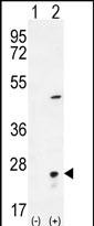
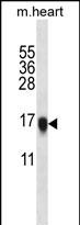
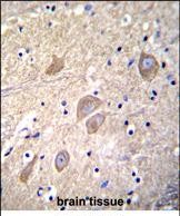
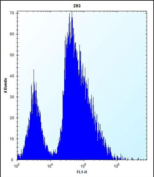
VSNL1 Antibody (Center)
Affinity Purified Rabbit Polyclonal Antibody (Pab)
- SPECIFICATION
- CITATIONS
- PROTOCOLS
- BACKGROUND

Application
| FC, IHC-P, WB, E |
|---|---|
| Primary Accession | P62760 |
| Other Accession | P62762, P62761, Q4R5F7, P62764, P62763, P35332, Q8BGZ1, Q9UM19, P29104, NP_003376 |
| Reactivity | Human, Mouse |
| Predicted | Bovine, Rat, Chicken, Monkey |
| Host | Rabbit |
| Clonality | Polyclonal |
| Isotype | Rabbit IgG |
| Calculated MW | 22142 Da |
| Antigen Region | 47-75 aa |
| Gene ID | 7447 |
|---|---|
| Other Names | Visinin-like protein 1, VILIP, VLP-1, Hippocalcin-like protein 3, HLP3, VSNL1, VISL1 |
| Target/Specificity | This VSNL1 antibody is generated from rabbits immunized with a KLH conjugated synthetic peptide between 47-75 amino acids from the Central region of human VSNL1. |
| Dilution | FC~~1:10~50 IHC-P~~1:10~50 WB~~1:1000 E~~Use at an assay dependent concentration. |
| Format | Purified polyclonal antibody supplied in PBS with 0.09% (W/V) sodium azide. This antibody is purified through a protein A column, followed by peptide affinity purification. |
| Storage | Maintain refrigerated at 2-8°C for up to 2 weeks. For long term storage store at -20°C in small aliquots to prevent freeze-thaw cycles. |
| Precautions | VSNL1 Antibody (Center) is for research use only and not for use in diagnostic or therapeutic procedures. |
| Name | VSNL1 |
|---|---|
| Synonyms | VISL1 |
| Function | Regulates (in vitro) the inhibition of rhodopsin phosphorylation in a calcium-dependent manner. |
| Tissue Location | Brain and retina. Neuron-specific in the central and peripheral nervous system. Increased in the cerebrospinal fluid of Alzheimer disease patients (at protein level) |

Thousands of laboratories across the world have published research that depended on the performance of antibodies from Abcepta to advance their research. Check out links to articles that cite our products in major peer-reviewed journals, organized by research category.
info@abcepta.com, and receive a free "I Love Antibodies" mug.

Provided below are standard protocols that you may find useful for product applications.
Background
This gene is a member of the visinin/recoverin subfamily of neuronal calcium sensor proteins. The encoded protein is strongly expressed in granule cells of the cerebellum where it associates with membranes in a calcium-dependent manner and modulates intracellular signaling pathways of the central nervous system by directly or indirectly regulating the activity of adenylyl cyclase. Alternatively spliced transcript variants have been observed, but their full-length nature has not been determined.
References
Xu, Y., et al. Schizophr. Res. 119 (1-3), 219-227 (2010) :
Buttgereit, J., et al. Regul. Pept. 161 (1-3), 51-57 (2010) :
Fu, J., et al. J. Biol. Chem. 284(40):27577-27586(2009)
Chen, K.C., et al. J. Pept. Sci. 15(2):89-94(2009)
Zhao, C.J., et al. Mol. Cell. Neurosci. 40(2):280-292(2009)
If you have used an Abcepta product and would like to share how it has performed, please click on the "Submit Review" button and provide the requested information. Our staff will examine and post your review and contact you if needed.
If you have any additional inquiries please email technical services at tech@abcepta.com.
Ordering Information
Other Products
Shipping Information
