


 Foundational characteristics of cancer include proliferation, angiogenesis, migration, evasion of apoptosis, and cellular immortality. Find key markers for these cellular processes and antibodies to detect them.
 The SUMOplot™ Analysis Program predicts and scores sumoylation sites in your protein. SUMOylation is a post-translational modification involved in various cellular processes, such as nuclear-cytosolic transport, transcriptional regulation, apoptosis, protein stability, response to stress, and progression through the cell cycle.
 The Autophagy Receptor Motif Plotter predicts and scores autophagy receptor binding sites in your protein. Identifying proteins connected to this pathway is critical to understanding the role of autophagy in physiological as well as pathological processes such as development, differentiation, neurodegenerative diseases, stress, infection, and cancer.
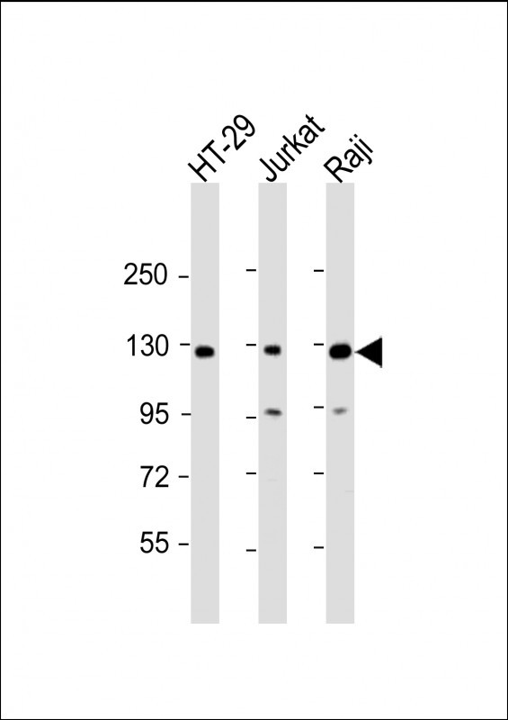
RBL2 Antibody (N-term)
Affinity Purified Rabbit Polyclonal Antibody (Pab)
- SPECIFICATION
- CITATIONS: 1
- PROTOCOLS
- BACKGROUND

Application
| FC, WB, E |
|---|---|
| Primary Accession | Q08999 |
| Other Accession | O55081, Q64700, NP_005602.3 |
| Reactivity | Human |
| Predicted | Mouse, Rat |
| Host | Rabbit |
| Clonality | Polyclonal |
| Isotype | Rabbit IgG |
| Calculated MW | 128367 Da |
| Antigen Region | 165-194 aa |
| Gene ID | 5934 |
|---|---|
| Other Names | Retinoblastoma-like protein 2, 130 kDa retinoblastoma-associated protein, p130, Retinoblastoma-related protein 2, RBR-2, pRb2, RBL2, RB2 |
| Target/Specificity | This RBL2 antibody is generated from rabbits immunized with a KLH conjugated synthetic peptide between 165-194 amino acids from the N-terminal region of human RBL2. |
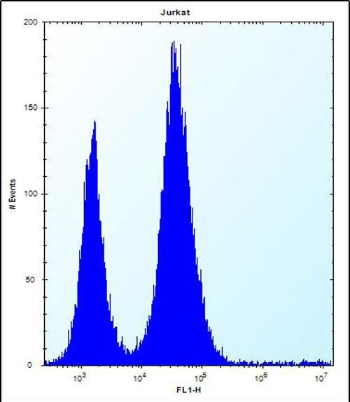
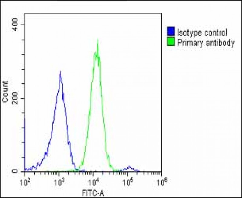
| Dilution | FC~~1:25 WB~~1:2000 E~~Use at an assay dependent concentration. |
| Format | Purified polyclonal antibody supplied in PBS with 0.09% (W/V) sodium azide. This antibody is purified through a protein A column, followed by peptide affinity purification. |
| Storage | Maintain refrigerated at 2-8°C for up to 2 weeks. For long term storage store at -20°C in small aliquots to prevent freeze-thaw cycles. |
| Precautions | RBL2 Antibody (N-term) is for research use only and not for use in diagnostic or therapeutic procedures. |
| Name | RBL2 |
|---|---|
| Synonyms | RB2 |
| Function | Key regulator of entry into cell division. Directly involved in heterochromatin formation by maintaining overall chromatin structure and, in particular, that of constitutive heterochromatin by stabilizing histone methylation. Recruits and targets histone methyltransferases KMT5B and KMT5C, leading to epigenetic transcriptional repression. Controls histone H4 'Lys-20' trimethylation. Probably acts as a transcription repressor by recruiting chromatin-modifying enzymes to promoters. Potent inhibitor of E2F-mediated trans-activation, associates preferentially with E2F5. Binds to cyclins A and E. Binds to and may be involved in the transforming capacity of the adenovirus E1A protein. May act as a tumor suppressor. |
| Cellular Location | Nucleus. |

Provided below are standard protocols that you may find useful for product applications.
Background
RBL2 is a key regulator of entry into cell division. Directly involved in heterochromatin formation by maintaining overall chromatin structure and, in particular, that of constitutive heterochromatin by stabilizing histone methylation. Recruits and targets histone methyltransferases SUV420H1 and SUV420H2, leading to epigenetic transcriptional repression. Controls histone H4 'Lys-20' trimethylation. Probably acts as a transcription repressor by recruiting chromatin-modifying enzymes to promoters. Potent inhibitor of E2F-mediated trans-activation, associates preferentially with E2F5. Binds to cyclins A and E. Binds to and may be involved in the transforming capacity of the adenovirus E1A protein. RBL2 may act as a tumor suppressor.
References
Schaffer, B.E., et al. Cancer Res. 70(10):3877-3883(2010)
Barrow-Laing, L., et al. Virology 400(2):233-239(2010)
Lu, F., et al. J. Virol. 84(6):2697-2706(2010)
Jowett, J.B., et al. Diabetes 59(3):726-732(2010)
Cunningham, J.M., et al. Br. J. Cancer 101(8):1461-1468(2009)
If you have used an Abcepta product and would like to share how it has performed, please click on the "Submit Review" button and provide the requested information. Our staff will examine and post your review and contact you if needed.
If you have any additional inquiries please email technical services at tech@abcepta.com.
Ordering Information
Other Products
Shipping Information
