


 Foundational characteristics of cancer include proliferation, angiogenesis, migration, evasion of apoptosis, and cellular immortality. Find key markers for these cellular processes and antibodies to detect them.
 The SUMOplot™ Analysis Program predicts and scores sumoylation sites in your protein. SUMOylation is a post-translational modification involved in various cellular processes, such as nuclear-cytosolic transport, transcriptional regulation, apoptosis, protein stability, response to stress, and progression through the cell cycle.
 The Autophagy Receptor Motif Plotter predicts and scores autophagy receptor binding sites in your protein. Identifying proteins connected to this pathway is critical to understanding the role of autophagy in physiological as well as pathological processes such as development, differentiation, neurodegenerative diseases, stress, infection, and cancer.
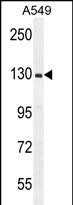
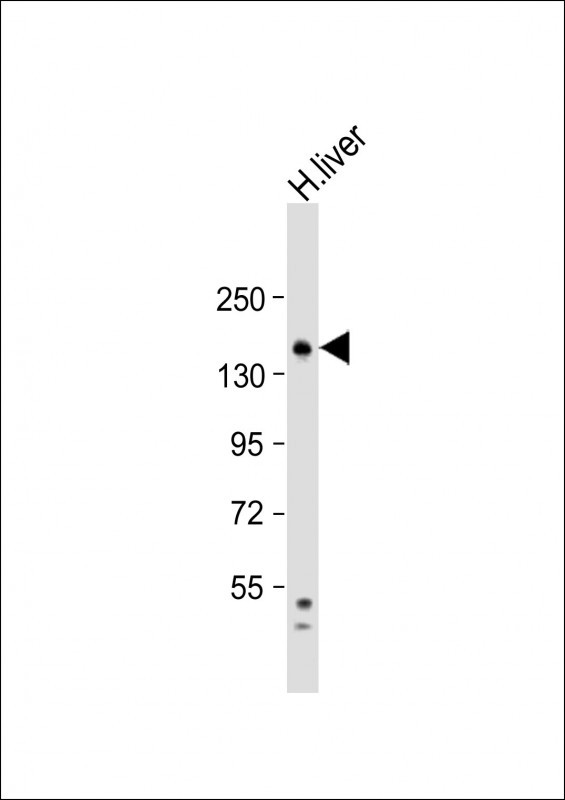
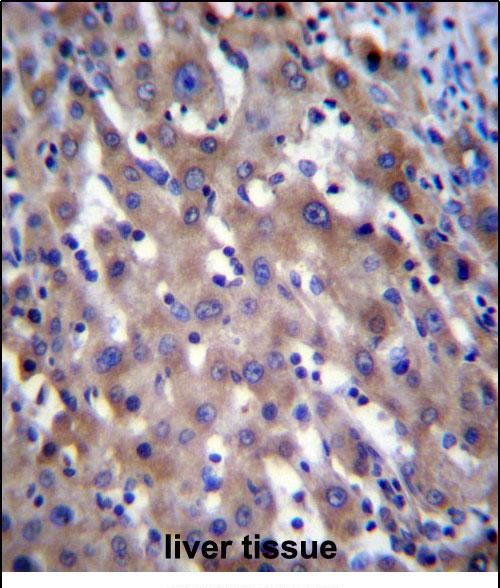
RPGR Antibody (C-term)
Affinity Purified Rabbit Polyclonal Antibody (Pab)
- SPECIFICATION
- CITATIONS
- PROTOCOLS
- BACKGROUND

Application
| IHC-P, WB, E |
|---|---|
| Primary Accession | Q92834 |
| Other Accession | NP_000319.1 |
| Reactivity | Human |
| Host | Rabbit |
| Clonality | Polyclonal |
| Isotype | Rabbit IgG |
| Calculated MW | 113387 Da |
| Antigen Region | 744-772 aa |
| Gene ID | 6103 |
|---|---|
| Other Names | X-linked retinitis pigmentosa GTPase regulator, RPGR, RP3, XLRP3 |
| Target/Specificity | This RPGR antibody is generated from rabbits immunized with a KLH conjugated synthetic peptide between 744-772 amino acids of human RPGR. |
| Dilution | IHC-P~~1:10~50 WB~~1:1000 E~~Use at an assay dependent concentration. |
| Format | Purified polyclonal antibody supplied in PBS with 0.09% (W/V) sodium azide. This antibody is purified through a protein A column, followed by peptide affinity purification. |
| Storage | Maintain refrigerated at 2-8°C for up to 2 weeks. For long term storage store at -20°C in small aliquots to prevent freeze-thaw cycles. |
| Precautions | RPGR Antibody (C-term) is for research use only and not for use in diagnostic or therapeutic procedures. |
| Name | RPGR (HGNC:10295) |
|---|---|
| Synonyms | RP3, XLRP3 |
| Function | Acts as a guanine-nucleotide releasing factor (GEF) for RAB8A and RAB37 by promoting the conversion of inactive RAB-GDP to the active form RAB-GTP (PubMed:20631154). GEF activity towards RAB8A may facilitate ciliary trafficking by modulating ciliary intracellular localization of RAB8A (PubMed:20631154). GEF activity towards RAB37 maintains autophagic homeostasis and retinal function (By similarity). Involved in photoreceptor integrity (By similarity). May control cilia formation by regulating actin stress filaments and cell contractility. May be involved in microtubule organization and regulation of transport in primary cilia (PubMed:21933838). May play a critical role in spermatogenesis and in intraflagellar transport processes (By similarity). |
| Cellular Location | Cytoplasm, cytoskeleton, flagellum axoneme {ECO:0000250|UniProtKB:Q9R0X5}. Golgi apparatus. Cell projection, cilium {ECO:0000250|UniProtKB:Q9R0X5}. Note=In the retinal photoreceptor cell layer, localizes at the connecting cilium (By similarity). Colocalizes with WHRN in the photoreceptor connecting cilium (By similarity) Colocalizes with CEP290 in the photoreceptor connecting cilium (By similarity). Colocalizes with RPGRIP1 in the photoreceptor connecting cilium (By similarity). Colocalizes with RPGR at the primary cilia of epithelial cells (By similarity). {ECO:0000250|UniProtKB:Q9N1T2, ECO:0000250|UniProtKB:Q9R0X5} |
| Tissue Location | Heart, brain, placenta, lung, liver, muscle, kidney, retina, pancreas and fetal retinal pigment epithelium. Isoform 3 is found only in the retina. Colocalizes with RPGRIP1 in the outer segment of rod photoreceptors and cone outer segments |

Thousands of laboratories across the world have published research that depended on the performance of antibodies from Abcepta to advance their research. Check out links to articles that cite our products in major peer-reviewed journals, organized by research category.
info@abcepta.com, and receive a free "I Love Antibodies" mug.

Provided below are standard protocols that you may find useful for product applications.
Background
This gene encodes a protein with a series of six RCC1-like domains (RLDs), characteristic of the highly conserved guanine nucleotide exchange factors. The encoded protein is found in the Golgi body and interacts with RPGRIP1. This protein localizes to the outer segment of rod photoreceptors and is essential for their viability. Mutations in this gene have been associated with X-linked retinitis pigmentosa (XLRP). Multiple alternatively spliced transcript variants that encode different isoforms of this gene have been reported, but the full-length natures of only some have been determined.
References
Clark, G.R., et al. Ophthalmology 117(11):2169-2177(2010)
Schmid, F., et al. Invest. Ophthalmol. Vis. Sci. 51(3):1628-1635(2010)
Ji, Y., et al. Curr. Eye Res. 35(1):73-79(2010)
Sheng, X., et al. Mol. Vis. 16, 1620-1628 (2010) :
Murga-Zamalloa, C.A., et al. J. Genet. 88(4):399-407(2009)
If you have used an Abcepta product and would like to share how it has performed, please click on the "Submit Review" button and provide the requested information. Our staff will examine and post your review and contact you if needed.
If you have any additional inquiries please email technical services at tech@abcepta.com.
Ordering Information
Other Products
Shipping Information
