


 Foundational characteristics of cancer include proliferation, angiogenesis, migration, evasion of apoptosis, and cellular immortality. Find key markers for these cellular processes and antibodies to detect them.
 The SUMOplot™ Analysis Program predicts and scores sumoylation sites in your protein. SUMOylation is a post-translational modification involved in various cellular processes, such as nuclear-cytosolic transport, transcriptional regulation, apoptosis, protein stability, response to stress, and progression through the cell cycle.
 The Autophagy Receptor Motif Plotter predicts and scores autophagy receptor binding sites in your protein. Identifying proteins connected to this pathway is critical to understanding the role of autophagy in physiological as well as pathological processes such as development, differentiation, neurodegenerative diseases, stress, infection, and cancer.
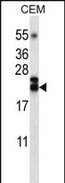
TNFSF4 Antibody (Center)
Affinity Purified Rabbit Polyclonal Antibody (Pab)
- SPECIFICATION
- CITATIONS
- PROTOCOLS
- BACKGROUND

Application
| WB, E |
|---|---|
| Primary Accession | P23510 |
| Other Accession | NP_003317.1 |
| Reactivity | Human |
| Host | Rabbit |
| Clonality | Polyclonal |
| Isotype | Rabbit IgG |
| Calculated MW | 21050 Da |
| Antigen Region | 102-131 aa |
| Gene ID | 7292 |
|---|---|
| Other Names | Tumor necrosis factor ligand superfamily member 4, Glycoprotein Gp34, OX40 ligand, OX40L, TAX transcriptionally-activated glycoprotein 1, CD252, TNFSF4, TXGP1 |
| Target/Specificity | This TNFSF4 antibody is generated from rabbits immunized with a KLH conjugated synthetic peptide between 102-131 amino acids from the Central region of human TNFSF4. |
| Dilution | WB~~1:1000 E~~Use at an assay dependent concentration. |
| Format | Purified polyclonal antibody supplied in PBS with 0.09% (W/V) sodium azide. This antibody is purified through a protein A column, followed by peptide affinity purification. |
| Storage | Maintain refrigerated at 2-8°C for up to 2 weeks. For long term storage store at -20°C in small aliquots to prevent freeze-thaw cycles. |
| Precautions | TNFSF4 Antibody (Center) is for research use only and not for use in diagnostic or therapeutic procedures. |
| Name | TNFSF4 |
|---|---|
| Synonyms | TXGP1 |
| Function | Cytokine that binds to TNFRSF4. Co-stimulates T-cell proliferation and cytokine production. |
| Cellular Location | Membrane; Single-pass type II membrane protein. |

Thousands of laboratories across the world have published research that depended on the performance of antibodies from Abcepta to advance their research. Check out links to articles that cite our products in major peer-reviewed journals, organized by research category.
info@abcepta.com, and receive a free "I Love Antibodies" mug.

Provided below are standard protocols that you may find useful for product applications.
Background
The protein encoded by this gene is a cytokine that belongs to the tumor necrosis factor (TNF) ligand family. This cytokine is a ligand for receptor TNFRSF4/OX4. It is found to be involved in T cell antigen-presenting cell (APC) interactions. In surface Ig- and CD40-stimulated B cells, this cytokine along with CD70 has been shown to provide CD28-independent costimulatory signals to T cells. This protein and its receptor are reported to directly mediate adhesion of activated T cells to vascular endothelial cells.
References
Karulf, M., et al. J. Immunol. 185(8):4856-4862(2010)
Shimada, M., et al. Hum. Genet. 128(4):433-441(2010)
Bailey, S.D., et al. Diabetes Care 33(10):2250-2253(2010)
Mosbruger, T.L., et al. J. Infect. Dis. 201(9):1371-1380(2010)
Davila, S., et al. Genes Immun. 11(3):232-238(2010)
If you have used an Abcepta product and would like to share how it has performed, please click on the "Submit Review" button and provide the requested information. Our staff will examine and post your review and contact you if needed.
If you have any additional inquiries please email technical services at tech@abcepta.com.
Ordering Information
Other Products
Shipping Information
