


 Foundational characteristics of cancer include proliferation, angiogenesis, migration, evasion of apoptosis, and cellular immortality. Find key markers for these cellular processes and antibodies to detect them.
 The SUMOplot™ Analysis Program predicts and scores sumoylation sites in your protein. SUMOylation is a post-translational modification involved in various cellular processes, such as nuclear-cytosolic transport, transcriptional regulation, apoptosis, protein stability, response to stress, and progression through the cell cycle.
 The Autophagy Receptor Motif Plotter predicts and scores autophagy receptor binding sites in your protein. Identifying proteins connected to this pathway is critical to understanding the role of autophagy in physiological as well as pathological processes such as development, differentiation, neurodegenerative diseases, stress, infection, and cancer.
TCF7L2 Antibody (N-term)
Affinity Purified Rabbit Polyclonal Antibody (Pab)
- SPECIFICATION
- CITATIONS
- PROTOCOLS
- BACKGROUND

Application
| WB, E |
|---|---|
| Primary Accession | Q9NQB0 |
| Other Accession | Q924A0, NP_001139756.1, NP_001139746.1 |
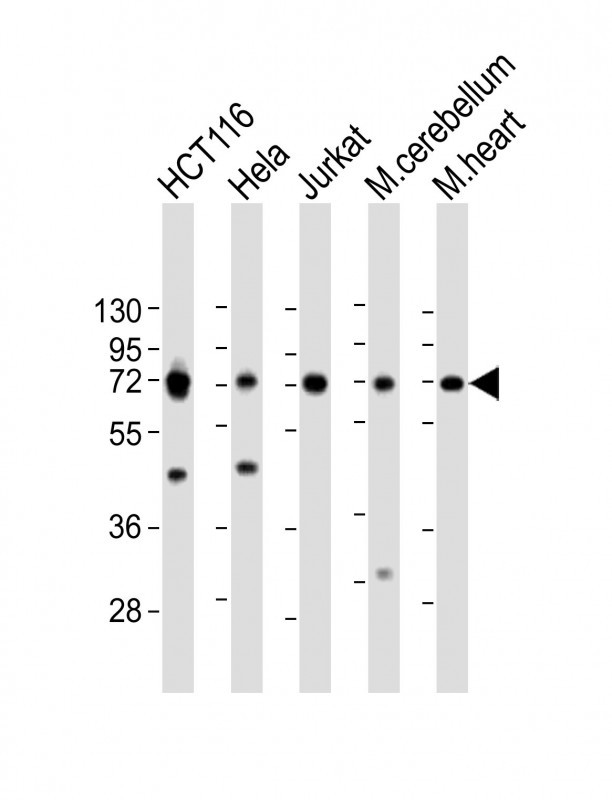
| Reactivity | Human, Mouse |
| Host | Rabbit |
| Clonality | Polyclonal |
| Isotype | Rabbit IgG |
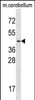
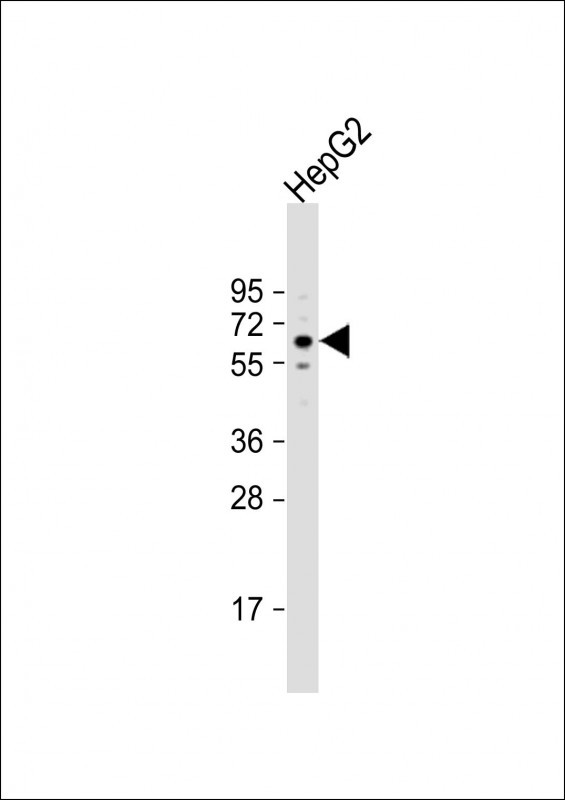
| Calculated MW | 67919 Da |
| Antigen Region | 61-90 aa |
| Gene ID | 6934 |
|---|---|
| Other Names | Transcription factor 7-like 2, HMG box transcription factor 4, T-cell-specific transcription factor 4, T-cell factor 4, TCF-4, hTCF-4, TCF7L2, TCF4 |
| Target/Specificity | This TCF7L2 antibody is generated from rabbits immunized with a KLH conjugated synthetic peptide between 61-90 amino acids from the N-terminal region of human TCF7L2. |
| Dilution | WB~~1:2000 E~~Use at an assay dependent concentration. |
| Format | Purified polyclonal antibody supplied in PBS with 0.09% (W/V) sodium azide. This antibody is purified through a protein A column, followed by peptide affinity purification. |
| Storage | Maintain refrigerated at 2-8°C for up to 2 weeks. For long term storage store at -20°C in small aliquots to prevent freeze-thaw cycles. |
| Precautions | TCF7L2 Antibody (N-term) is for research use only and not for use in diagnostic or therapeutic procedures. |
| Name | TCF7L2 |
|---|---|
| Synonyms | TCF4 |
| Function | Participates in the Wnt signaling pathway and modulates MYC expression by binding to its promoter in a sequence-specific manner. Acts as a repressor in the absence of CTNNB1, and as activator in its presence. Activates transcription from promoters with several copies of the Tcf motif 5'-CCTTTGATC-3' in the presence of CTNNB1. TLE1, TLE2, TLE3 and TLE4 repress transactivation mediated by TCF7L2/TCF4 and CTNNB1. Expression of dominant-negative mutants results in cell-cycle arrest in G1. Necessary for the maintenance of the epithelial stem-cell compartment of the small intestine. |
| Cellular Location | Nucleus, PML body. Nucleus. Note=Diffuse pattern. Colocalizes with SUMO1 and PIAS4 in a subset of PML (promyelocytic leukemia) nuclear bodies |
| Tissue Location | Detected in epithelium from small intestine, with the highest expression at the top of the crypts and a gradient of expression from crypt to villus. Detected in colon epithelium and colon cancer, and in epithelium from mammary gland and carcinomas derived therefrom. |

Thousands of laboratories across the world have published research that depended on the performance of antibodies from Abcepta to advance their research. Check out links to articles that cite our products in major peer-reviewed journals, organized by research category.
info@abcepta.com, and receive a free "I Love Antibodies" mug.

Provided below are standard protocols that you may find useful for product applications.
Background
This gene encodes a high mobility group (HMG) box-containing transcription factor that plays a key role in the Wnt signaling pathway. The protein has been implicated in blood glucose homeostasis. Genetic variants of this gene are associated with increased risk of type 2 diabetes. Several transcript variants encoding multiple different isoforms have been found for this gene.
References
Hansson, O., et al. Curr. Diab. Rep. 10(6):444-451(2010)
Heni, M., et al. Diabetes (2010) In press :
Potapov, V.A., et al. Genetika 46(8):1123-1131(2010)
Kucharska-Newton, A.M., et al. J Obes 2010 (2010) :
Zabaneh, D., et al. PLoS ONE 5 (8), E11961 (2010) :
If you have used an Abcepta product and would like to share how it has performed, please click on the "Submit Review" button and provide the requested information. Our staff will examine and post your review and contact you if needed.
If you have any additional inquiries please email technical services at tech@abcepta.com.
Ordering Information
Other Products
Shipping Information
