


 Foundational characteristics of cancer include proliferation, angiogenesis, migration, evasion of apoptosis, and cellular immortality. Find key markers for these cellular processes and antibodies to detect them.
 The SUMOplot™ Analysis Program predicts and scores sumoylation sites in your protein. SUMOylation is a post-translational modification involved in various cellular processes, such as nuclear-cytosolic transport, transcriptional regulation, apoptosis, protein stability, response to stress, and progression through the cell cycle.
 The Autophagy Receptor Motif Plotter predicts and scores autophagy receptor binding sites in your protein. Identifying proteins connected to this pathway is critical to understanding the role of autophagy in physiological as well as pathological processes such as development, differentiation, neurodegenerative diseases, stress, infection, and cancer.
PEX14 Antibody (Center)
Affinity Purified Rabbit Polyclonal Antibody (Pab)
- SPECIFICATION
- CITATIONS
- PROTOCOLS
- BACKGROUND

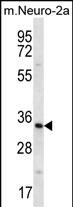
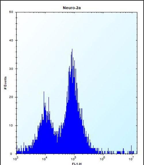
Application
| FC, WB, E |
|---|---|
| Primary Accession | O75381 |
| Other Accession | NP_004556.1 |
| Reactivity | Human, Mouse |
| Host | Rabbit |
| Clonality | Polyclonal |
| Isotype | Rabbit IgG |
| Calculated MW | 41237 Da |
| Antigen Region | 121-150 aa |
| Gene ID | 5195 |
|---|---|
| Other Names | Peroxisomal membrane protein PEX14, PTS1 receptor-docking protein, Peroxin-14, Peroxisomal membrane anchor protein PEX14, PEX14 |
| Target/Specificity | This PEX14 antibody is generated from rabbits immunized with a KLH conjugated synthetic peptide between 121-150 amino acids from the Central region of human PEX14. |
| Dilution | FC~~1:10~50 WB~~1:1000 E~~Use at an assay dependent concentration. |
| Format | Purified polyclonal antibody supplied in PBS with 0.09% (W/V) sodium azide. This antibody is purified through a protein A column, followed by peptide affinity purification. |
| Storage | Maintain refrigerated at 2-8°C for up to 2 weeks. For long term storage store at -20°C in small aliquots to prevent freeze-thaw cycles. |
| Precautions | PEX14 Antibody (Center) is for research use only and not for use in diagnostic or therapeutic procedures. |
| Name | PEX14 {ECO:0000303|PubMed:9653144, ECO:0000312|HGNC:HGNC:8856} |
|---|---|
| Function | Component of the PEX13-PEX14 docking complex, a translocon channel that specifically mediates the import of peroxisomal cargo proteins bound to PEX5 receptor (PubMed:24235149, PubMed:28765278, PubMed:9653144). The PEX13-PEX14 docking complex forms a large import pore which can be opened to a diameter of about 9 nm (By similarity). Mechanistically, PEX5 receptor along with cargo proteins associates with the PEX14 subunit of the PEX13-PEX14 docking complex in the cytosol, leading to the insertion of the receptor into the organelle membrane with the concomitant translocation of the cargo into the peroxisome matrix (PubMed:24235149, PubMed:28765278). Plays a key role for peroxisome movement through a direct interaction with tubulin (PubMed:21525035). |
| Cellular Location | Peroxisome membrane; Single-pass membrane protein {ECO:0000250|UniProtKB:Q642G4} |

Thousands of laboratories across the world have published research that depended on the performance of antibodies from Abcepta to advance their research. Check out links to articles that cite our products in major peer-reviewed journals, organized by research category.
info@abcepta.com, and receive a free "I Love Antibodies" mug.

Provided below are standard protocols that you may find useful for product applications.
Background
This gene encodes an essential component of the peroxisomal import machinery. The protein is integrated into peroxisome membranes with its C-terminus exposed to the cytosol, and interacts with the cytosolic receptor for proteins containing a PTS1 peroxisomal targeting signal. The protein also functions as a transcriptional corepressor and interacts with a histone deacetylase. A mutation in this gene results in one form of Zellweger syndrome.
References
Bailey, S.D., et al. Diabetes Care 33(10):2250-2253(2010) Macgregor, S., et al. Hum. Mol. Genet. 19(13):2716-2724(2010) Rose, J.E., et al. Mol. Med. 16 (7-8), 247-253 (2010) : Talmud, P.J., et al. Am. J. Hum. Genet. 85(5):628-642(2009) Shiozawa, K., et al. J. Biol. Chem. 284(37):25334-25342(2009)
If you have used an Abcepta product and would like to share how it has performed, please click on the "Submit Review" button and provide the requested information. Our staff will examine and post your review and contact you if needed.
If you have any additional inquiries please email technical services at tech@abcepta.com.
Ordering Information
Other Products
Shipping Information
