


 Foundational characteristics of cancer include proliferation, angiogenesis, migration, evasion of apoptosis, and cellular immortality. Find key markers for these cellular processes and antibodies to detect them.
 The SUMOplot™ Analysis Program predicts and scores sumoylation sites in your protein. SUMOylation is a post-translational modification involved in various cellular processes, such as nuclear-cytosolic transport, transcriptional regulation, apoptosis, protein stability, response to stress, and progression through the cell cycle.
 The Autophagy Receptor Motif Plotter predicts and scores autophagy receptor binding sites in your protein. Identifying proteins connected to this pathway is critical to understanding the role of autophagy in physiological as well as pathological processes such as development, differentiation, neurodegenerative diseases, stress, infection, and cancer.
CLEC3B Antibody (Center)
Purified Rabbit Polyclonal Antibody (Pab)
- SPECIFICATION
- CITATIONS: 1
- PROTOCOLS
- BACKGROUND

Application
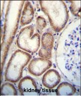
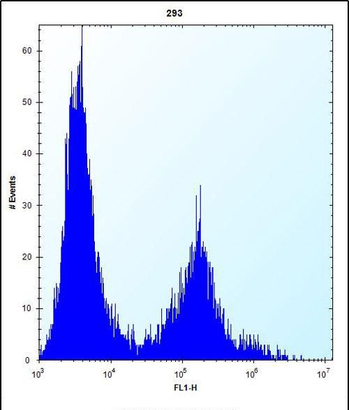
| FC, IHC-P, WB, E |
|---|---|
| Primary Accession | P05452 |
| Other Accession | Q2KIS7, NP_003269.2 |
| Reactivity | Human |
| Predicted | Bovine |
| Host | Rabbit |
| Clonality | Polyclonal |
| Isotype | Rabbit IgG |
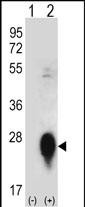
| Calculated MW | 22537 Da |
| Antigen Region | 95-122 aa |
| Gene ID | 7123 |
|---|---|
| Other Names | Tetranectin, TN, C-type lectin domain family 3 member B, Plasminogen kringle 4-binding protein, CLEC3B, TNA |
| Target/Specificity | This CLEC3B antibody is generated from rabbits immunized with a KLH conjugated synthetic peptide between 95-122 amino acids from the Central region of human CLEC3B. |
| Dilution | FC~~1:10~50 IHC-P~~1:10~50 WB~~1:1000 E~~Use at an assay dependent concentration. |
| Format | Purified polyclonal antibody supplied in PBS with 0.09% (W/V) sodium azide. This antibody is prepared by Saturated Ammonium Sulfate (SAS) precipitation followed by dialysis against PBS. |
| Storage | Maintain refrigerated at 2-8°C for up to 2 weeks. For long term storage store at -20°C in small aliquots to prevent freeze-thaw cycles. |
| Precautions | CLEC3B Antibody (Center) is for research use only and not for use in diagnostic or therapeutic procedures. |
| Name | CLEC3B |
|---|---|
| Synonyms | TNA |
| Function | Tetranectin binds to plasminogen and to isolated kringle 4. May be involved in the packaging of molecules destined for exocytosis. Plays a role in retinal function (PubMed:35331648). |
| Cellular Location | Secreted. |
| Tissue Location | Found in plasma. |

Provided below are standard protocols that you may find useful for product applications.
Background
Tetranectin binds to plasminogen and to isolated kringle 4. It may be involved in the packaging of molecules destined for exocytosis.
References
Ewing, R.M., et al. Mol. Syst. Biol. 3, 89 (2007) :
Valdes, A.M., et al. Arthritis Rheum. 54(2):533-539(2006)
Hermann, M., et al. Transplant. Proc. 37(2):1322-1325(2005)
Anderson, N.L., et al. Mol. Cell Proteomics 3(4):311-326(2004)
Westergaard, U.B., et al. Eur. J. Biochem. 270(8):1850-1854(2003)
If you have used an Abcepta product and would like to share how it has performed, please click on the "Submit Review" button and provide the requested information. Our staff will examine and post your review and contact you if needed.
If you have any additional inquiries please email technical services at tech@abcepta.com.
Ordering Information
Other Products
Shipping Information
