


 Foundational characteristics of cancer include proliferation, angiogenesis, migration, evasion of apoptosis, and cellular immortality. Find key markers for these cellular processes and antibodies to detect them.
 The SUMOplot™ Analysis Program predicts and scores sumoylation sites in your protein. SUMOylation is a post-translational modification involved in various cellular processes, such as nuclear-cytosolic transport, transcriptional regulation, apoptosis, protein stability, response to stress, and progression through the cell cycle.
 The Autophagy Receptor Motif Plotter predicts and scores autophagy receptor binding sites in your protein. Identifying proteins connected to this pathway is critical to understanding the role of autophagy in physiological as well as pathological processes such as development, differentiation, neurodegenerative diseases, stress, infection, and cancer.
MN1 Antibody (Center)
Affinity Purified Rabbit Polyclonal Antibody (Pab)
- SPECIFICATION
- CITATIONS
- PROTOCOLS
- BACKGROUND

Application
| WB, E |
|---|---|
| Primary Accession | Q10571 |
| Other Accession | NP_002421.3 |
| Reactivity | Human |
| Host | Rabbit |
| Clonality | Polyclonal |
| Isotype | Rabbit IgG |
| Calculated MW | 136001 Da |
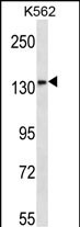
| Antigen Region | 835-864 aa |
| Gene ID | 4330 |
|---|---|
| Other Names | Probable tumor suppressor protein MN1, MN1 |
| Target/Specificity | This MN1 antibody is generated from rabbits immunized with a KLH conjugated synthetic peptide between 835-864 amino acids from the Central region of human MN1. |
| Dilution | WB~~1:1000 E~~Use at an assay dependent concentration. |
| Format | Purified polyclonal antibody supplied in PBS with 0.09% (W/V) sodium azide. This antibody is purified through a protein A column, followed by peptide affinity purification. |
| Storage | Maintain refrigerated at 2-8°C for up to 2 weeks. For long term storage store at -20°C in small aliquots to prevent freeze-thaw cycles. |
| Precautions | MN1 Antibody (Center) is for research use only and not for use in diagnostic or therapeutic procedures. |
| Name | MN1 |
|---|---|
| Function | Transcriptional activator which specifically regulates expression of TBX22 in the posterior region of the developing palate. Required during later stages of palate development for growth and medial fusion of the palatal shelves. Promotes maturation and normal function of calvarial osteoblasts, including expression of the osteoclastogenic cytokine TNFSF11/RANKL. Necessary for normal development of the membranous bones of the skull (By similarity). May play a role in tumor suppression (Probable). |
| Cellular Location | Nucleus. |
| Tissue Location | Widely expressed in fetal and adult tissues. Highest expression is observed in fetal brain and skeletal muscle, and adult skeletal muscle. |

Thousands of laboratories across the world have published research that depended on the performance of antibodies from Abcepta to advance their research. Check out links to articles that cite our products in major peer-reviewed journals, organized by research category.
info@abcepta.com, and receive a free "I Love Antibodies" mug.

Provided below are standard protocols that you may find useful for product applications.
Background
Meningioma 1 (MN1) contains two sets of CAG repeats. It is disrupted by a balanced translocation (4;22) in a meningioma, and its inactivation may contribute to meningioma 32 pathogenesis.
References
Liu, T., et al. Leukemia 24(3):601-612(2010)
Kandilci, A., et al. Blood 114(8):1596-1606(2009)
Trynka, G., et al. Gut 58(8):1078-1083(2009)
Langer, C., et al. J. Clin. Oncol. 27(19):3198-3204(2009)
Schroeder, T., et al. Leuk. Lymphoma 50(6):1043-1046(2009)
If you have used an Abcepta product and would like to share how it has performed, please click on the "Submit Review" button and provide the requested information. Our staff will examine and post your review and contact you if needed.
If you have any additional inquiries please email technical services at tech@abcepta.com.
Ordering Information
Other Products
Shipping Information
