


 Foundational characteristics of cancer include proliferation, angiogenesis, migration, evasion of apoptosis, and cellular immortality. Find key markers for these cellular processes and antibodies to detect them.
 The SUMOplot™ Analysis Program predicts and scores sumoylation sites in your protein. SUMOylation is a post-translational modification involved in various cellular processes, such as nuclear-cytosolic transport, transcriptional regulation, apoptosis, protein stability, response to stress, and progression through the cell cycle.
 The Autophagy Receptor Motif Plotter predicts and scores autophagy receptor binding sites in your protein. Identifying proteins connected to this pathway is critical to understanding the role of autophagy in physiological as well as pathological processes such as development, differentiation, neurodegenerative diseases, stress, infection, and cancer.
XYLT1 Antibody (N-term)
Affinity Purified Rabbit Polyclonal Antibody (Pab)
- SPECIFICATION
- CITATIONS
- PROTOCOLS
- BACKGROUND

Application
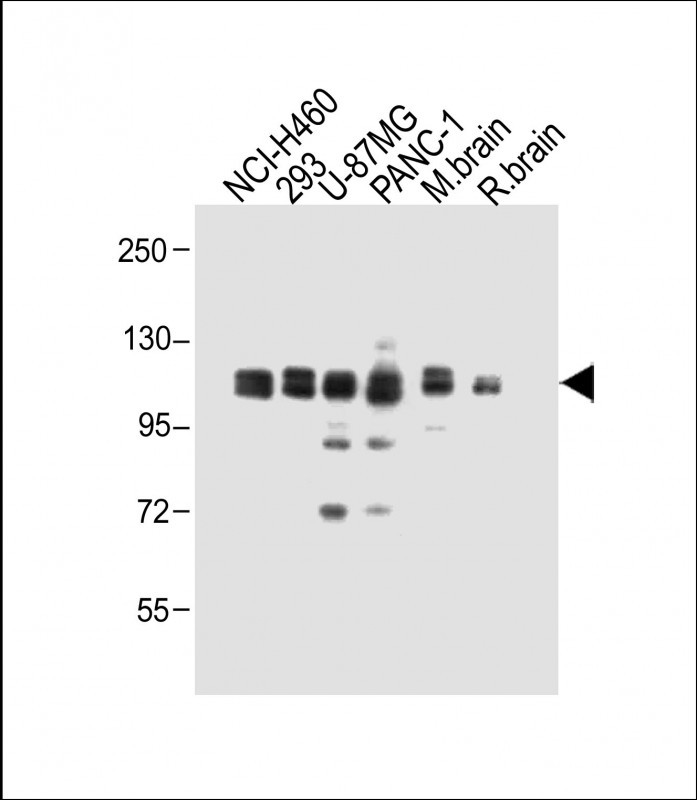
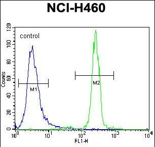
| WB, FC, E |
|---|---|
| Primary Accession | Q86Y38 |
| Other Accession | Q9EPI1, Q811B1, NP_071449.1 |
| Reactivity | Human, Mouse, Rat |
| Predicted | Mouse, Rat |
| Host | Rabbit |
| Clonality | Polyclonal |
| Isotype | Rabbit IgG |
| Calculated MW | 107569 Da |
| Antigen Region | 126-155 aa |
| Gene ID | 64131 |
|---|---|
| Other Names | Xylosyltransferase 1, Peptide O-xylosyltransferase 1, Xylosyltransferase I, XT-I, XylT-I, XYLT1, XT1 |
| Target/Specificity | This XYLT1 antibody is generated from rabbits immunized with a KLH conjugated synthetic peptide between 126-155 amino acids from the N-terminal region of human XYLT1. |
| Dilution | WB~~1:1000 FC~~1:10~50 E~~Use at an assay dependent concentration. |
| Format | Purified polyclonal antibody supplied in PBS with 0.09% (W/V) sodium azide. This antibody is purified through a protein A column, followed by peptide affinity purification. |
| Storage | Maintain refrigerated at 2-8°C for up to 2 weeks. For long term storage store at -20°C in small aliquots to prevent freeze-thaw cycles. |
| Precautions | XYLT1 Antibody (N-term) is for research use only and not for use in diagnostic or therapeutic procedures. |
| Name | XYLT1 |
|---|---|
| Synonyms | XT1 |
| Function | Catalyzes the first step in the biosynthesis of chondroitin sulfate and dermatan sulfate proteoglycans, such as DCN. Transfers D- xylose from UDP-D-xylose to specific serine residues of the core protein (PubMed:15461586, PubMed:17189265, PubMed:23982343, PubMed:24581741). Required for normal embryonic and postnatal skeleton development, especially of the long bones (PubMed:23982343, PubMed:24581741). Required for normal maturation of chondrocytes during bone development, and normal onset of ossification (By similarity). |
| Cellular Location | Golgi apparatus membrane; Single-pass type II membrane protein. Secreted Note=Detected predominantly in the Golgi apparatus |
| Tissue Location | Widely expressed. Expressed at higher level in placenta, kidney and pancreas. Weakly expressed in skeletal muscle |

Thousands of laboratories across the world have published research that depended on the performance of antibodies from Abcepta to advance their research. Check out links to articles that cite our products in major peer-reviewed journals, organized by research category.
info@abcepta.com, and receive a free "I Love Antibodies" mug.

Provided below are standard protocols that you may find useful for product applications.
Background
This locus encodes a xylosyltransferase enzyme. The encoded protein catalyzes transfer of UDP-xylose to serine residues of an acceptor protein substrate. This transfer reaction is necessary for biosynthesis of glycosaminoglycan chains. Mutations in this gene have been associated with increased severity of pseudoxanthoma elasticum.
References
Rose, J.E., et al. Mol. Med. 16 (7-8), 247-253 (2010) :
Muller, B., et al. J. Biol. Chem. 284(45):30775-30782(2009)
Ponighaus, C., et al. Am. J. Hypertens. 22(4):432-436(2009)
Ambrosius, M., et al. Clin. Biochem. 42 (1-2), 1-4 (2009) :
Schon, S., et al. J. Med. Genet. 43(9):745-749(2006)
If you have used an Abcepta product and would like to share how it has performed, please click on the "Submit Review" button and provide the requested information. Our staff will examine and post your review and contact you if needed.
If you have any additional inquiries please email technical services at tech@abcepta.com.
Ordering Information
Other Products
Shipping Information
