


 Foundational characteristics of cancer include proliferation, angiogenesis, migration, evasion of apoptosis, and cellular immortality. Find key markers for these cellular processes and antibodies to detect them.
 The SUMOplot™ Analysis Program predicts and scores sumoylation sites in your protein. SUMOylation is a post-translational modification involved in various cellular processes, such as nuclear-cytosolic transport, transcriptional regulation, apoptosis, protein stability, response to stress, and progression through the cell cycle.
 The Autophagy Receptor Motif Plotter predicts and scores autophagy receptor binding sites in your protein. Identifying proteins connected to this pathway is critical to understanding the role of autophagy in physiological as well as pathological processes such as development, differentiation, neurodegenerative diseases, stress, infection, and cancer.
DULLARD Antibody (Center)
Affinity Purified Rabbit Polyclonal Antibody (Pab)

- SPECIFICATION
- CITATIONS
- PROTOCOLS
- BACKGROUND

Application
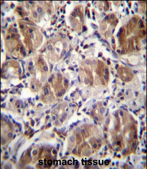
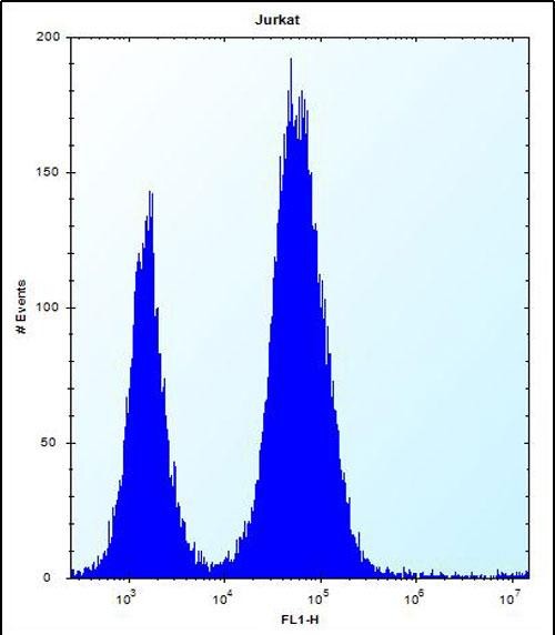
| WB, FC, IHC-P, E |
|---|---|
| Primary Accession | O95476 |
| Other Accession | Q3B7T6, Q3TP92, Q1RMV9, NP_001137247.1 |
| Reactivity | Human |
| Predicted | Bovine, Mouse, Rat |
| Host | Rabbit |
| Clonality | Polyclonal |
| Isotype | Rabbit IgG |
| Calculated MW | 28377 Da |
| Antigen Region | 131-160 aa |
| Gene ID | 23399 |
|---|---|
| Other Names | CTD nuclear envelope phosphatase 1, Serine/threonine-protein phosphatase dullard, CTDNEP1, DULLARD |
| Target/Specificity | This DULLARD antibody is generated from rabbits immunized with a KLH conjugated synthetic peptide between 131-160 amino acids from the Central region of human DULLARD. |
| Dilution | WB~~1:1000 FC~~1:10~50 IHC-P~~1:10~50 E~~Use at an assay dependent concentration. |
| Format | Purified polyclonal antibody supplied in PBS with 0.09% (W/V) sodium azide. This antibody is purified through a protein A column, followed by peptide affinity purification. |
| Storage | Maintain refrigerated at 2-8°C for up to 2 weeks. For long term storage store at -20°C in small aliquots to prevent freeze-thaw cycles. |
| Precautions | DULLARD Antibody (Center) is for research use only and not for use in diagnostic or therapeutic procedures. |
| Name | CTDNEP1 |
|---|---|
| Synonyms | DULLARD |
| Function | Serine/threonine protein phosphatase forming with CNEP1R1 an active phosphatase complex that dephosphorylates and may activate LPIN1 and LPIN2. LPIN1 and LPIN2 are phosphatidate phosphatases that catalyze the conversion of phosphatidic acid to diacylglycerol and control the metabolism of fatty acids at different levels. May indirectly modulate the lipid composition of nuclear and/or endoplasmic reticulum membranes and be required for proper nuclear membrane morphology and/or dynamics. May also indirectly regulate the production of lipid droplets and triacylglycerol. May antagonize BMP signaling. |
| Cellular Location | Endoplasmic reticulum membrane; Single-pass membrane protein. Nucleus membrane; Single-pass membrane protein |
| Tissue Location | Muscle specific with lower expression in other metabolic tissues. |

Thousands of laboratories across the world have published research that depended on the performance of antibodies from Abcepta to advance their research. Check out links to articles that cite our products in major peer-reviewed journals, organized by research category.
info@abcepta.com, and receive a free "I Love Antibodies" mug.

Provided below are standard protocols that you may find useful for product applications.
Background
Serine/threonine phosphatase which may be required for proper nuclear membrane morphology. Involved in LPIN1 dephosphorylation. May antagonize BMP signaling.
References
Kim, Y., et al. Proc. Natl. Acad. Sci. U.S.A. 104(16):6596-6601(2007)
Zhang, Y., et al. Mol. Cell 24(5):759-770(2006)
Satow, R., et al. Dev. Cell 11(6):763-774(2006)
Satow, R., et al. Biochem. Biophys. Res. Commun. 295(1):85-91(2002)
If you have used an Abcepta product and would like to share how it has performed, please click on the "Submit Review" button and provide the requested information. Our staff will examine and post your review and contact you if needed.
If you have any additional inquiries please email technical services at tech@abcepta.com.
Ordering Information
Other Products
Shipping Information
