


 Foundational characteristics of cancer include proliferation, angiogenesis, migration, evasion of apoptosis, and cellular immortality. Find key markers for these cellular processes and antibodies to detect them.
 The SUMOplot™ Analysis Program predicts and scores sumoylation sites in your protein. SUMOylation is a post-translational modification involved in various cellular processes, such as nuclear-cytosolic transport, transcriptional regulation, apoptosis, protein stability, response to stress, and progression through the cell cycle.
 The Autophagy Receptor Motif Plotter predicts and scores autophagy receptor binding sites in your protein. Identifying proteins connected to this pathway is critical to understanding the role of autophagy in physiological as well as pathological processes such as development, differentiation, neurodegenerative diseases, stress, infection, and cancer.
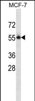
PVRL3 Antibody (C-term)
Affinity Purified Rabbit Polyclonal Antibody (Pab)
- SPECIFICATION
- CITATIONS
- PROTOCOLS
- BACKGROUND

Application
| WB, E |
|---|---|
| Primary Accession | Q9NQS3 |
| Other Accession | NP_056295.1 |
| Reactivity | Human |
| Host | Rabbit |
| Clonality | Polyclonal |
| Isotype | Rabbit IgG |
| Calculated MW | 61002 Da |
| Antigen Region | 507-536 aa |
| Gene ID | 25945 |
|---|---|
| Other Names | Nectin-3, CDw113, Poliovirus receptor-related protein 3, CD113, PVRL3, PRR3 |
| Target/Specificity | This PVRL3 antibody is generated from rabbits immunized with a KLH conjugated synthetic peptide between 507-536 amino acids from the C-terminal region of human PVRL3. |
| Dilution | WB~~1:1000 E~~Use at an assay dependent concentration. |
| Format | Purified polyclonal antibody supplied in PBS with 0.09% (W/V) sodium azide. This antibody is purified through a protein A column, followed by peptide affinity purification. |
| Storage | Maintain refrigerated at 2-8°C for up to 2 weeks. For long term storage store at -20°C in small aliquots to prevent freeze-thaw cycles. |
| Precautions | PVRL3 Antibody (C-term) is for research use only and not for use in diagnostic or therapeutic procedures. |
| Name | NECTIN3 (HGNC:17664) |
|---|---|
| Synonyms | PRR3, PVRL3 |
| Function | Cell adhesion molecule that promotes cell-cell adhesion through heterophilic trans-interactions with nectins-like or other nectins, such as trans-interaction with NECTIN2 at Sertoli-spermatid junctions (PubMed:16216929). Trans-interaction with PVR induces activation of CDC42 and RAC small G proteins through common signaling molecules such as SRC and RAP1 (PubMed:16216929). Induces endocytosis- mediated down-regulation of PVR from the cell surface, resulting in reduction of cell movement and proliferation (PubMed:16216929). Involved in axon guidance by promoting contacts between the commissural axons and the floor plate cells (By similarity). Also involved in the formation of cell-cell junctions, including adherens junctions and synapses (By similarity). Promotes formation of checkerboard-like cellular pattern of hair cells and supporting cells in the auditory epithelium via heterophilic interaction with NECTIN1: NECTIN1 is present in the membrane of hair cells and associates with NECTIN3 on supporting cells, thereby mediating heterotypic adhesion between these two cell types (By similarity). Plays a role in the morphology of the ciliary body (By similarity). |
| Cellular Location | Cell membrane; Single-pass membrane protein. Postsynaptic cell membrane {ECO:0000250|UniProtKB:Q9JLB9}; Single-pass type I membrane protein. Cell junction, adherens junction {ECO:0000250|UniProtKB:Q9JLB9}. Note=In the auditory epithelium, specificaly localizes to the apical side of the lateral membranes of supporting cells. {ECO:0000250|UniProtKB:Q9JLB9} |
| Tissue Location | Predominantly expressed in testis and placenta as well as in many cell lines, including epithelial cell lines |

Thousands of laboratories across the world have published research that depended on the performance of antibodies from Abcepta to advance their research. Check out links to articles that cite our products in major peer-reviewed journals, organized by research category.
info@abcepta.com, and receive a free "I Love Antibodies" mug.

Provided below are standard protocols that you may find useful for product applications.
Background
Nectins (e.g., PVRL1; MIM 600644) are immunoglobulin-like adhesion molecules that interact with afadin (AF6; MIM 159559). Afadin is an actin filament-binding protein that connects nectins to the actin cytoskeleton. The nectin-afadin system organizes adherens junctions cooperatively with the cadherin (see MIM 192090)-catenin (see MIM 116805) system in epithelial cells.
References
Bailey, S.D., et al. Diabetes Care 33(10):2250-2253(2010)
Jugessur, A., et al. PLoS ONE 5 (7), E11493 (2010) :
Talmud, P.J., et al. Am. J. Hum. Genet. 85(5):628-642(2009)
Yu, X., et al. Nat. Immunol. 10(1):48-57(2009)
Fujito, T., et al. J. Cell Biol. 171(1):165-173(2005)
If you have used an Abcepta product and would like to share how it has performed, please click on the "Submit Review" button and provide the requested information. Our staff will examine and post your review and contact you if needed.
If you have any additional inquiries please email technical services at tech@abcepta.com.
Ordering Information
Other Products
Shipping Information
