


 Foundational characteristics of cancer include proliferation, angiogenesis, migration, evasion of apoptosis, and cellular immortality. Find key markers for these cellular processes and antibodies to detect them.
 The SUMOplot™ Analysis Program predicts and scores sumoylation sites in your protein. SUMOylation is a post-translational modification involved in various cellular processes, such as nuclear-cytosolic transport, transcriptional regulation, apoptosis, protein stability, response to stress, and progression through the cell cycle.
 The Autophagy Receptor Motif Plotter predicts and scores autophagy receptor binding sites in your protein. Identifying proteins connected to this pathway is critical to understanding the role of autophagy in physiological as well as pathological processes such as development, differentiation, neurodegenerative diseases, stress, infection, and cancer.
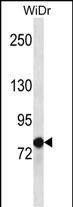
LZTS2 Antibody (C-term)
Affinity Purified Rabbit Polyclonal Antibody (Pab)
- SPECIFICATION
- CITATIONS
- PROTOCOLS
- BACKGROUND

Application
| WB, E |
|---|---|
| Primary Accession | Q9BRK4 |
| Other Accession | NP_115805.1 |
| Reactivity | Human |
| Host | Rabbit |
| Clonality | Polyclonal |
| Isotype | Rabbit IgG |
| Calculated MW | 72759 Da |
| Antigen Region | 572-601 aa |
| Gene ID | 84445 |
|---|---|
| Other Names | Leucine zipper putative tumor suppressor 2 {ECO:0000255|HAMAP-Rule:MF_03026}, hLZTS2, Protein LAPSER1 {ECO:0000255|HAMAP-Rule:MF_03026}, LZTS2 {ECO:0000255|HAMAP-Rule:MF_03026} |
| Target/Specificity | This LZTS2 antibody is generated from rabbits immunized with a KLH conjugated synthetic peptide between 572-601 amino acids from the C-terminal region of human LZTS2. |
| Dilution | WB~~1:1000 E~~Use at an assay dependent concentration. |
| Format | Purified polyclonal antibody supplied in PBS with 0.09% (W/V) sodium azide. This antibody is purified through a protein A column, followed by peptide affinity purification. |
| Storage | Maintain refrigerated at 2-8°C for up to 2 weeks. For long term storage store at -20°C in small aliquots to prevent freeze-thaw cycles. |
| Precautions | LZTS2 Antibody (C-term) is for research use only and not for use in diagnostic or therapeutic procedures. |
| Name | LZTS2 {ECO:0000255|HAMAP-Rule:MF_03026} |
|---|---|
| Function | Negative regulator of katanin-mediated microtubule severing and release from the centrosome. Required for central spindle formation and the completion of cytokinesis. May negatively regulate axonal outgrowth by preventing the formation of microtubule bundles that are necessary for transport within the elongating axon. Negative regulator of the Wnt signaling pathway. Represses beta-catenin-mediated transcriptional activation by promoting the nuclear exclusion of beta- catenin. |
| Cellular Location | Cytoplasm. Cytoplasm, cytoskeleton, microtubule organizing center, centrosome. Note=Localized to the centrosome throughout the cell cycle. Localized to the midbody in cells undergoing cytokinesis |
| Tissue Location | Highly expressed in prostate and testis, and at slightly lower levels in spleen, thymus, uterus, small intestine and colon. |

Thousands of laboratories across the world have published research that depended on the performance of antibodies from Abcepta to advance their research. Check out links to articles that cite our products in major peer-reviewed journals, organized by research category.
info@abcepta.com, and receive a free "I Love Antibodies" mug.

Provided below are standard protocols that you may find useful for product applications.
Background
Negative regulator of katanin-mediated microtubule severing and release from the centrosome. Required for central spindle formation and the completion of cytokinesis. May negatively regulate axonal outgrowth by preventing the formation of microtubule bundles that are necessary for transport within the elongating axon. Negative regulator of the Wnt signaling pathway. Represses beta-catenin-mediated transcriptional activation by promoting the nuclear exclusion of beta-catenin.
References
Olson, J.E., et al. Breast Cancer Res. Treat. (2010) In press :
Sudo, H., et al. Hum. Mol. Genet. 17(16):2524-2540(2008)
Hyun Hwa, Cho., et al. Biochim. Biophys. Acta 1783(3):419-428(2008)
Lim, J., et al. Cell 125(4):801-814(2006)
Grupe, A., et al. Am. J. Hum. Genet. 78(1):78-88(2006)
If you have used an Abcepta product and would like to share how it has performed, please click on the "Submit Review" button and provide the requested information. Our staff will examine and post your review and contact you if needed.
If you have any additional inquiries please email technical services at tech@abcepta.com.
Ordering Information
Shipping Information
