


 Foundational characteristics of cancer include proliferation, angiogenesis, migration, evasion of apoptosis, and cellular immortality. Find key markers for these cellular processes and antibodies to detect them.
 The SUMOplot™ Analysis Program predicts and scores sumoylation sites in your protein. SUMOylation is a post-translational modification involved in various cellular processes, such as nuclear-cytosolic transport, transcriptional regulation, apoptosis, protein stability, response to stress, and progression through the cell cycle.
 The Autophagy Receptor Motif Plotter predicts and scores autophagy receptor binding sites in your protein. Identifying proteins connected to this pathway is critical to understanding the role of autophagy in physiological as well as pathological processes such as development, differentiation, neurodegenerative diseases, stress, infection, and cancer.
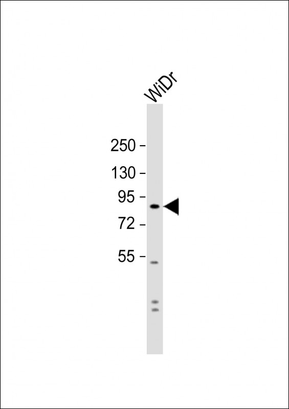
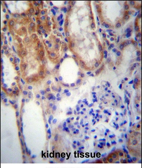
SUSD2 Antibody (C-term)
Affinity Purified Rabbit Polyclonal Antibody (Pab)
- SPECIFICATION
- CITATIONS
- PROTOCOLS
- BACKGROUND

Application
| IHC-P, WB, E |
|---|---|
| Primary Accession | Q9UGT4 |
| Other Accession | NP_062547.1 |
| Reactivity | Human |
| Host | Rabbit |
| Clonality | Polyclonal |
| Isotype | Rabbit IgG |
| Calculated MW | 90208 Da |
| Antigen Region | 716-745 aa |
| Gene ID | 56241 |
|---|---|
| Other Names | Sushi domain-containing protein 2, SUSD2 |
| Target/Specificity | This SUSD2 antibody is generated from rabbits immunized with a KLH conjugated synthetic peptide between 716-745 amino acids from the C-terminal region of human SUSD2. |
| Dilution | IHC-P~~1:10~50 WB~~1:1000 E~~Use at an assay dependent concentration. |
| Format | Purified polyclonal antibody supplied in PBS with 0.09% (W/V) sodium azide. This antibody is purified through a protein A column, followed by peptide affinity purification. |
| Storage | Maintain refrigerated at 2-8°C for up to 2 weeks. For long term storage store at -20°C in small aliquots to prevent freeze-thaw cycles. |
| Precautions | SUSD2 Antibody (C-term) is for research use only and not for use in diagnostic or therapeutic procedures. |
| Name | SUSD2 |
|---|---|
| Function | May be a cytokine receptor for GPR15LG. May be a tumor suppressor; together with GPR15LG has a growth inhibitory effect on colon cancer cells which includes G1 cell cycle arrest (PubMed:25351403). May play a role in breast tumorigenesis (PubMed:23131994). |
| Cellular Location | Cell membrane; Single-pass type I membrane protein. Note=SUSD2 and LGALS1 co-localized in very specific, punctate regions along the cell membrane of breast cancer cells |
| Tissue Location | Highly expressed in breast cancer, but shows a restricted expression pattern in normal tissues such as adipose, adrenal gland, kidney, lung, mammary gland, placenta, thyroid, trachea, and uterus (PubMed:23131994). Also expressed in colon; down-regulated in colon cancer tissues (PubMed:25351403) |

Thousands of laboratories across the world have published research that depended on the performance of antibodies from Abcepta to advance their research. Check out links to articles that cite our products in major peer-reviewed journals, organized by research category.
info@abcepta.com, and receive a free "I Love Antibodies" mug.

Provided below are standard protocols that you may find useful for product applications.
References
Ewing, R.M., et al. Mol. Syst. Biol. 3, 89 (2007) :
Liu, T., et al. J. Proteome Res. 4(6):2070-2080(2005)
Dunham, I., et al. Nature 402(6761):489-495(1999)
If you have used an Abcepta product and would like to share how it has performed, please click on the "Submit Review" button and provide the requested information. Our staff will examine and post your review and contact you if needed.
If you have any additional inquiries please email technical services at tech@abcepta.com.
Ordering Information
Other Products
Shipping Information
