


 Foundational characteristics of cancer include proliferation, angiogenesis, migration, evasion of apoptosis, and cellular immortality. Find key markers for these cellular processes and antibodies to detect them.
 The SUMOplot™ Analysis Program predicts and scores sumoylation sites in your protein. SUMOylation is a post-translational modification involved in various cellular processes, such as nuclear-cytosolic transport, transcriptional regulation, apoptosis, protein stability, response to stress, and progression through the cell cycle.
 The Autophagy Receptor Motif Plotter predicts and scores autophagy receptor binding sites in your protein. Identifying proteins connected to this pathway is critical to understanding the role of autophagy in physiological as well as pathological processes such as development, differentiation, neurodegenerative diseases, stress, infection, and cancer.
MPP3 Antibody (C-term)
Affinity Purified Rabbit Polyclonal Antibody (Pab)
- SPECIFICATION
- CITATIONS
- PROTOCOLS
- BACKGROUND

Application
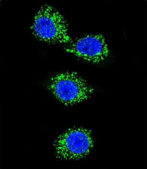
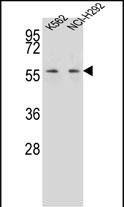
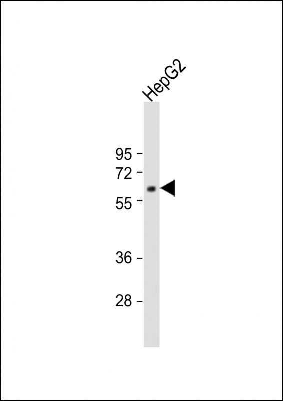
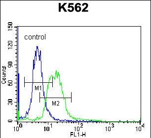
| FC, IF, WB, E |
|---|---|
| Primary Accession | Q13368 |
| Other Accession | NP_001923.2 |
| Reactivity | Human |
| Host | Rabbit |
| Clonality | Polyclonal |
| Isotype | Rabbit IgG |
| Calculated MW | 66152 Da |
| Antigen Region | 406-434 aa |
| Gene ID | 4356 |
|---|---|
| Other Names | MAGUK p55 subfamily member 3, Discs large homolog 3, Protein MPP3, MPP3, DLG3 |
| Target/Specificity | This MPP3 antibody is generated from rabbits immunized with a KLH conjugated synthetic peptide between 406-434 amino acids from the C-terminal region of human MPP3. |
| Dilution | FC~~1:10~50 IF~~1:10~50 WB~~1:1000 E~~Use at an assay dependent concentration. |
| Format | Purified polyclonal antibody supplied in PBS with 0.09% (W/V) sodium azide. This antibody is purified through a protein A column, followed by peptide affinity purification. |
| Storage | Maintain refrigerated at 2-8°C for up to 2 weeks. For long term storage store at -20°C in small aliquots to prevent freeze-thaw cycles. |
| Precautions | MPP3 Antibody (C-term) is for research use only and not for use in diagnostic or therapeutic procedures. |
| Name | MPP3 {ECO:0000303|PubMed:16519681, ECO:0000312|HGNC:HGNC:7221} |
|---|---|
| Function | Participates in cell spreading through the phosphoinositide- 3-kinase (PI3K) pathway by connecting CADM1 to DLG1 and the regulatory subunit of phosphoinositide-3-kinase (PI3K) (PubMed:24503895). Stabilizes HTR2C at the plasma membrane and prevents its desensitization. May participates in the maintenance of adherens junctions (By similarity). |
| Cellular Location | Cell membrane. Apical cell membrane. Cell junction, adherens junction. Note=Localized in apical villi of Mueller glia cells (By similarity). Localized at the apical membrane in the developing cortex and colocalized with apical proteins and adherens junction proteins (By similarity). Localized at the outer limiting membrane (OLM), and outer plexiform (OPL) of retina (PubMed:16519681). {ECO:0000250|UniProtKB:O88910, ECO:0000269|PubMed:16519681} |
| Tissue Location | Expressed in retina (at protein level) at the subapical region (SAR) adjacent to adherens junctions at the OLM, and at the OPL. |

Thousands of laboratories across the world have published research that depended on the performance of antibodies from Abcepta to advance their research. Check out links to articles that cite our products in major peer-reviewed journals, organized by research category.
info@abcepta.com, and receive a free "I Love Antibodies" mug.

Provided below are standard protocols that you may find useful for product applications.
Background
This gene product is a member of a family of membrane-associated proteins termed MAGUKs (membrane-associated guanylate kinase homologs). MAGUKs interact with the cytoskeleton and regulate cell proliferation, signaling pathways, and intracellular junctions. This protein contains a conserved sequence, called the SH3 (src homology 3) motif, found in several other proteins that associate with the cytoskeleton and are suspected to play important roles in signal transduction. Alternatively spliced transcript variants have been identified. One transcript variant is experimentally supported, but it doesn't encode a protein.
References
Qin, L., et al. Zhonghua Gan Zang Bing Za Zhi 15(7):509-512(2007)
Kantardzhieva, A., et al. FEBS J. 273(6):1152-1165(2006)
Stelzl, U., et al. Cell 122(6):957-968(2005)
Fukuhara, H., et al. Oncogene 22(40):6160-6165(2003)
Shin, B.K., et al. J. Biol. Chem. 278(9):7607-7616(2003)
If you have used an Abcepta product and would like to share how it has performed, please click on the "Submit Review" button and provide the requested information. Our staff will examine and post your review and contact you if needed.
If you have any additional inquiries please email technical services at tech@abcepta.com.
Ordering Information
Shipping Information
