


 Foundational characteristics of cancer include proliferation, angiogenesis, migration, evasion of apoptosis, and cellular immortality. Find key markers for these cellular processes and antibodies to detect them.
 The SUMOplot™ Analysis Program predicts and scores sumoylation sites in your protein. SUMOylation is a post-translational modification involved in various cellular processes, such as nuclear-cytosolic transport, transcriptional regulation, apoptosis, protein stability, response to stress, and progression through the cell cycle.
 The Autophagy Receptor Motif Plotter predicts and scores autophagy receptor binding sites in your protein. Identifying proteins connected to this pathway is critical to understanding the role of autophagy in physiological as well as pathological processes such as development, differentiation, neurodegenerative diseases, stress, infection, and cancer.
MCL1 Antibody (BH3 Domain Specific)
Purified Rabbit Polyclonal Antibody (Pab)
- SPECIFICATION
- CITATIONS: 4
- PROTOCOLS
- BACKGROUND

Application
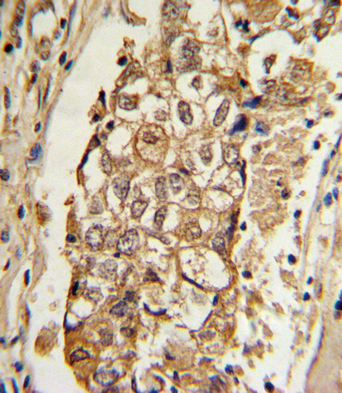
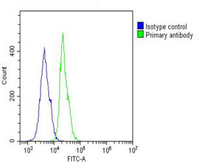
| IHC-P, WB, FC, E |
|---|---|
| Primary Accession | Q07820 |
| Reactivity | Human, Mouse, Rat |
| Host | Rabbit |
| Clonality | Polyclonal |
| Isotype | Rabbit IgG |
| Calculated MW | 37337 Da |
| Antigen Region | 191-226 aa |
| Gene ID | 4170 |
|---|---|
| Other Names | Induced myeloid leukemia cell differentiation protein Mcl-1, Bcl-2-like protein 3, Bcl2-L-3, Bcl-2-related protein EAT/mcl1, mcl1/EAT, MCL1, BCL2L3 |
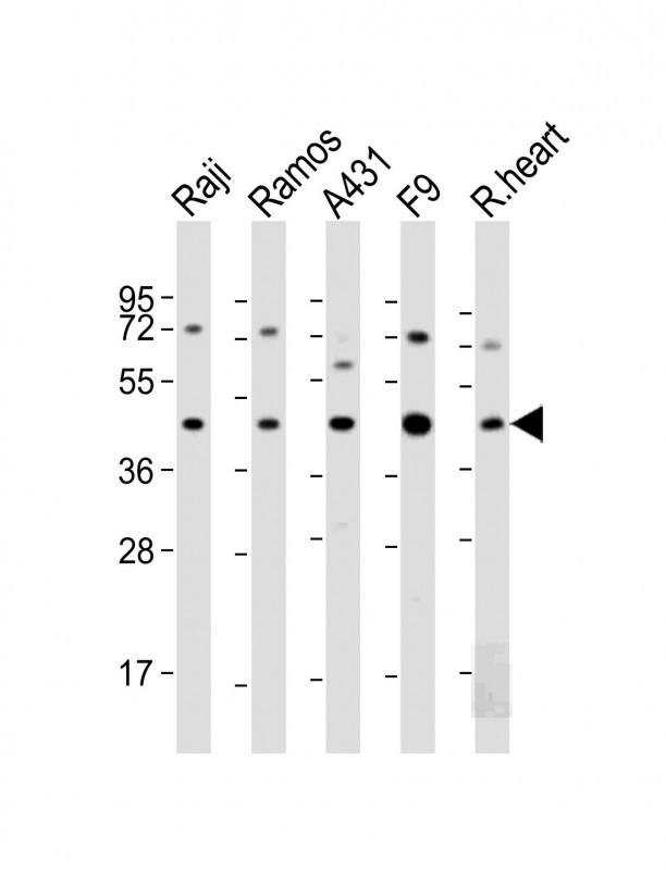
| Target/Specificity | This MCL1 antibody is generated from rabbits immunized with a KLH conjugated synthetic peptide between 191-226 amino acids from human MCL1. |
| Dilution | IHC-P~~1:10~50 WB~~1:2000 FC~~1:25 E~~Use at an assay dependent concentration. |
| Format | Purified polyclonal antibody supplied in PBS with 0.09% (W/V) sodium azide. This antibody is purified through a protein A column, followed by peptide affinity purification. |
| Storage | Maintain refrigerated at 2-8°C for up to 2 weeks. For long term storage store at -20°C in small aliquots to prevent freeze-thaw cycles. |
| Precautions | MCL1 Antibody (BH3 Domain Specific) is for research use only and not for use in diagnostic or therapeutic procedures. |
| Name | MCL1 |
|---|---|
| Synonyms | BCL2L3 |
| Function | Involved in the regulation of apoptosis versus cell survival, and in the maintenance of viability but not of proliferation. Mediates its effects by interactions with a number of other regulators of apoptosis. Isoform 1 inhibits apoptosis. Isoform 2 promotes apoptosis. |
| Cellular Location | Membrane; Single-pass membrane protein. Cytoplasm. Mitochondrion. Nucleus, nucleoplasm Note=Cytoplasmic, associated with mitochondria |

Provided below are standard protocols that you may find useful for product applications.
Background
The Mcl-1 protein belongs to the Bcl-2 family. Alternative splicing occurs at this locus and two transcript variants encoding distinct isoforms have been identified. The longer gene product (isoform 1) enhances cell survival by inhibiting apoptosis while the alternatively spliced shorter gene product (isoform 2) promotes apoptosis and is death-inducing.
References
Crossley, L.J., J. Leukoc. Biol. 74(4):583-592 (2003).
Kotelkin, A., et al., J. Virol. 77(17):9156-9172 (2003).
Erwert, R.D., et al., Microb. Pathog. 35(2):87-93 (2003).
Liu, H., et al., Blood 102(1):344-352 (2003).
Nijhawan, D., et al., Genes Dev. 17(12):1475-1486 (2003).
If you have used an Abcepta product and would like to share how it has performed, please click on the "Submit Review" button and provide the requested information. Our staff will examine and post your review and contact you if needed.
If you have any additional inquiries please email technical services at tech@abcepta.com.
Ordering Information
Other Products
Shipping Information
