


 Foundational characteristics of cancer include proliferation, angiogenesis, migration, evasion of apoptosis, and cellular immortality. Find key markers for these cellular processes and antibodies to detect them.
 The SUMOplot™ Analysis Program predicts and scores sumoylation sites in your protein. SUMOylation is a post-translational modification involved in various cellular processes, such as nuclear-cytosolic transport, transcriptional regulation, apoptosis, protein stability, response to stress, and progression through the cell cycle.
 The Autophagy Receptor Motif Plotter predicts and scores autophagy receptor binding sites in your protein. Identifying proteins connected to this pathway is critical to understanding the role of autophagy in physiological as well as pathological processes such as development, differentiation, neurodegenerative diseases, stress, infection, and cancer.
REPS2 Antibody (N-term)
Affinity Purified Rabbit Polyclonal Antibody (Pab)
- SPECIFICATION
- CITATIONS
- PROTOCOLS
- BACKGROUND

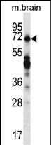
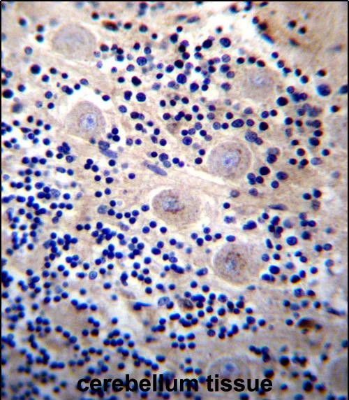
Application
| IHC-P, WB, E |
|---|---|
| Primary Accession | Q8NFH8 |
| Other Accession | NP_004717.2, NP_001074444.1 |
| Reactivity | Human, Mouse |
| Host | Rabbit |
| Clonality | Polyclonal |
| Isotype | Rabbit IgG |
| Calculated MW | 71534 Da |
| Antigen Region | 153-182 aa |
| Gene ID | 9185 |
|---|---|
| Other Names | RalBP1-associated Eps domain-containing protein 2, Partner of RalBP1, RalBP1-interacting protein 2, REPS2, POB1 |
| Target/Specificity | This REPS2 antibody is generated from rabbits immunized with a KLH conjugated synthetic peptide between 153-182 amino acids from the N-terminal region of human REPS2. |
| Dilution | IHC-P~~1:10~50 WB~~1:1000 E~~Use at an assay dependent concentration. |
| Format | Purified polyclonal antibody supplied in PBS with 0.09% (W/V) sodium azide. This antibody is purified through a protein A column, followed by peptide affinity purification. |
| Storage | Maintain refrigerated at 2-8°C for up to 2 weeks. For long term storage store at -20°C in small aliquots to prevent freeze-thaw cycles. |
| Precautions | REPS2 Antibody (N-term) is for research use only and not for use in diagnostic or therapeutic procedures. |
| Name | REPS2 |
|---|---|
| Synonyms | POB1 |
| Function | Involved in ligand-dependent receptor mediated endocytosis of the EGF and insulin receptors as part of the Ral signaling pathway (PubMed:10393179, PubMed:12771942, PubMed:9422736). By controlling growth factor receptors endocytosis may regulate cell survival (PubMed:12771942). Through ASAP1 may regulate cell adhesion and migration (PubMed:12149250). |
| Cellular Location | Cytoplasm. |
| Tissue Location | Expressed at high levels in the cerebrum, cerebellum, lung, kidney, and testis. Weakly expressed in the kidney Isoform 2 is down-regulated during progression of prostate cancer |

Thousands of laboratories across the world have published research that depended on the performance of antibodies from Abcepta to advance their research. Check out links to articles that cite our products in major peer-reviewed journals, organized by research category.
info@abcepta.com, and receive a free "I Love Antibodies" mug.

Provided below are standard protocols that you may find useful for product applications.
Background
The product of this gene is part of a protein complex that regulates the endocytosis of growth factor receptors. The encoded protein directly interacts with a GTPase activating protein that functions downstream of the small G protein Ral. Its expression can negatively affect receptor internalization and inhibit growth factor signaling. Multiple transcript variants encoding different isoforms have been found for this gene.
References
Doolan, P., et al. Tumour Biol. 30(4):200-209(2009) Singhal, S.S., et al. J. Biol. Chem. 283(28):19714-19729(2008) Yadav, S., et al. Biochem. Biophys. Res. Commun. 328(4):1003-1009(2005) Oosterhoff, J.K., et al. Int. J. Cancer 113(4):561-567(2005) Penninkhof, F., et al. Oncogene 23(33):5607-5615(2004)
If you have used an Abcepta product and would like to share how it has performed, please click on the "Submit Review" button and provide the requested information. Our staff will examine and post your review and contact you if needed.
If you have any additional inquiries please email technical services at tech@abcepta.com.
Ordering Information
Shipping Information
