


 Foundational characteristics of cancer include proliferation, angiogenesis, migration, evasion of apoptosis, and cellular immortality. Find key markers for these cellular processes and antibodies to detect them.
 The SUMOplot™ Analysis Program predicts and scores sumoylation sites in your protein. SUMOylation is a post-translational modification involved in various cellular processes, such as nuclear-cytosolic transport, transcriptional regulation, apoptosis, protein stability, response to stress, and progression through the cell cycle.
 The Autophagy Receptor Motif Plotter predicts and scores autophagy receptor binding sites in your protein. Identifying proteins connected to this pathway is critical to understanding the role of autophagy in physiological as well as pathological processes such as development, differentiation, neurodegenerative diseases, stress, infection, and cancer.
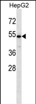
CHST9 Antibody (N-term)
Affinity Purified Rabbit Polyclonal Antibody (Pab)
- SPECIFICATION
- CITATIONS
- PROTOCOLS
- BACKGROUND

Application
| WB, E |
|---|---|
| Primary Accession | Q7L1S5 |
| Other Accession | NP_113610.2 |
| Reactivity | Human |
| Host | Rabbit |
| Clonality | Polyclonal |
| Isotype | Rabbit IgG |
| Calculated MW | 52055 Da |
| Antigen Region | 21-50 aa |
| Gene ID | 83539 |
|---|---|
| Other Names | Carbohydrate sulfotransferase 9, 282-, GalNAc-4-O-sulfotransferase 2, GalNAc-4-ST2, GalNAc4ST-2, N-acetylgalactosamine-4-O-sulfotransferase 2, CHST9 |
| Target/Specificity | This CHST9 antibody is generated from rabbits immunized with a KLH conjugated synthetic peptide between 21-50 amino acids from the N-terminal region of human CHST9. |
| Dilution | WB~~1:1000 E~~Use at an assay dependent concentration. |
| Format | Purified polyclonal antibody supplied in PBS with 0.09% (W/V) sodium azide. This antibody is purified through a protein A column, followed by peptide affinity purification. |
| Storage | Maintain refrigerated at 2-8°C for up to 2 weeks. For long term storage store at -20°C in small aliquots to prevent freeze-thaw cycles. |
| Precautions | CHST9 Antibody (N-term) is for research use only and not for use in diagnostic or therapeutic procedures. |
| Name | CHST9 |
|---|---|
| Function | Catalyzes the transfer of sulfate to position 4 of non- reducing N-acetylgalactosamine (GalNAc) residues in both N-glycans and O-glycans. Participates in biosynthesis of glycoprotein hormones lutropin and thyrotropin, by mediating sulfation of their carbohydrate structures. Has a higher activity toward carbonic anhydrase VI than toward lutropin. Only active against terminal GalNAcbeta1,GalNAcbeta. Isoform 2, but not isoform 1, is active toward chondroitin. |
| Cellular Location | [Isoform 1]: Golgi apparatus membrane; Single-pass type II membrane protein |
| Tissue Location | Highly expressed in trachea. Also expressed in fetal lung, adult pancreas, testis and salivary gland. Expressed at low level in pituitary gland, apex of the heart, adult lung, prostate and mammary gland. Weakly or not expressed in heart, liver and spinal cord |

Thousands of laboratories across the world have published research that depended on the performance of antibodies from Abcepta to advance their research. Check out links to articles that cite our products in major peer-reviewed journals, organized by research category.
info@abcepta.com, and receive a free "I Love Antibodies" mug.

Provided below are standard protocols that you may find useful for product applications.
Background
Sulfate groups in carbohydrates confer highly specific functions on glycoproteins, glycolipids, and proteoglycans and are critical for cell-cell interaction, signal transduction, and embryonic development. Sulfotransferases, such as CHST9, carry out sulfation of carbohydrates (Hiraoka et al., 2001 [PubMed 11445554]).
References
Clark, H.F., et al. Genome Res. 13(10):2265-2270(2003)
Hiraoka, N., et al. Glycobiology 11(6):495-504(2001)
Kang, H.G., et al. J. Biol. Chem. 276(14):10861-10869(2001)
If you have used an Abcepta product and would like to share how it has performed, please click on the "Submit Review" button and provide the requested information. Our staff will examine and post your review and contact you if needed.
If you have any additional inquiries please email technical services at tech@abcepta.com.
Ordering Information
Other Products
Shipping Information
