


 Foundational characteristics of cancer include proliferation, angiogenesis, migration, evasion of apoptosis, and cellular immortality. Find key markers for these cellular processes and antibodies to detect them.
 The SUMOplot™ Analysis Program predicts and scores sumoylation sites in your protein. SUMOylation is a post-translational modification involved in various cellular processes, such as nuclear-cytosolic transport, transcriptional regulation, apoptosis, protein stability, response to stress, and progression through the cell cycle.
 The Autophagy Receptor Motif Plotter predicts and scores autophagy receptor binding sites in your protein. Identifying proteins connected to this pathway is critical to understanding the role of autophagy in physiological as well as pathological processes such as development, differentiation, neurodegenerative diseases, stress, infection, and cancer.
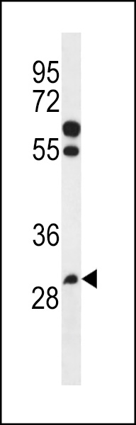
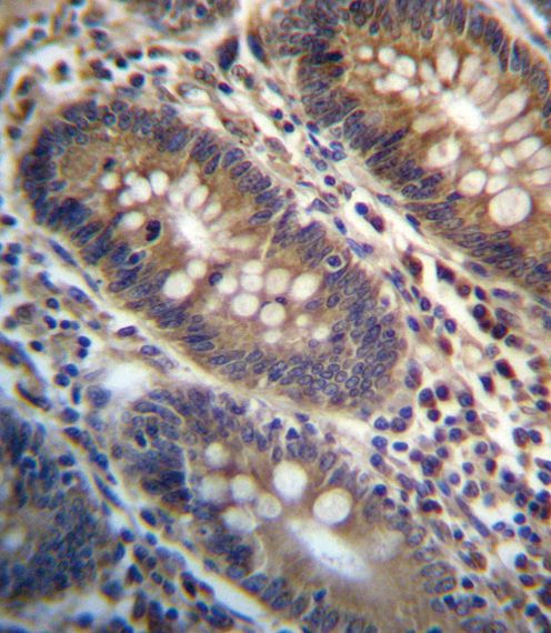
PGAP3 Antibody (Center)
Affinity Purified Rabbit Polyclonal Antibody (Pab)
- SPECIFICATION
- CITATIONS
- PROTOCOLS
- BACKGROUND

Application
| IHC-P, WB, E |
|---|---|
| Primary Accession | Q96FM1 |
| Other Accession | A2A559, A2V7M9, A7YWP2, NP_219487.3 |
| Reactivity | Human |
| Predicted | Bovine, Hamster, Mouse |
| Host | Rabbit |
| Clonality | Polyclonal |
| Isotype | Rabbit IgG |
| Calculated MW | 36475 Da |
| Antigen Region | 141-169 aa |
| Gene ID | 93210 |
|---|---|
| Other Names | Post-GPI attachment to proteins factor 3, COS16 homolog, hCOS16, Gene coamplified with ERBB2 protein, PER1-like domain-containing protein 1, PGAP3, CAB2, PERLD1 |
| Target/Specificity | This PGAP3 antibody is generated from rabbits immunized with a KLH conjugated synthetic peptide between 141-169 amino acids from the Central region of human PGAP3. |
| Dilution | IHC-P~~1:10~50 WB~~1:1000 E~~Use at an assay dependent concentration. |
| Format | Purified polyclonal antibody supplied in PBS with 0.09% (W/V) sodium azide. This antibody is purified through a protein A column, followed by peptide affinity purification. |
| Storage | Maintain refrigerated at 2-8°C for up to 2 weeks. For long term storage store at -20°C in small aliquots to prevent freeze-thaw cycles. |
| Precautions | PGAP3 Antibody (Center) is for research use only and not for use in diagnostic or therapeutic procedures. |
| Name | PGAP3 (HGNC:23719) |
|---|---|
| Synonyms | CAB2, PERLD1 |
| Function | Involved in the fatty acid remodeling steps of GPI-anchor maturation where the unsaturated acyl chain at sn-2 of inositol phosphate is replaced by a saturated stearoyl chain (PubMed:17021251, PubMed:24439110). May catalyze the first step of the fatty acid remodeling, by removing the unsaturated acyl chain at sn-2 of inositol phosphate, generating a lyso-GPI intermediate (Probable). The fatty acid remodeling steps is critical for the integration of GPI-APs into lipid rafts (By similarity). |
| Cellular Location | Golgi apparatus membrane; Multi-pass membrane protein |
| Tissue Location | Ubiquitously expressed, with highest levels in thyroid and placenta. |

Thousands of laboratories across the world have published research that depended on the performance of antibodies from Abcepta to advance their research. Check out links to articles that cite our products in major peer-reviewed journals, organized by research category.
info@abcepta.com, and receive a free "I Love Antibodies" mug.

Provided below are standard protocols that you may find useful for product applications.
Background
PGAP3 is involved in the lipid remodeling steps of GPI-anchor maturation. Lipid remodeling steps consist in the generation of 2 saturated fatty chains at the sn-2 position of GPI-anchors proteins. Required for phospholipase A2 activity that removes an acyl-chain at the sn-2 position of GPI-anchors during the remodeling of GPI (Probable).
References
Bailey, S.D., et al. Diabetes Care (2010) In press :
Talmud, P.J., et al. Am. J. Hum. Genet. 85(5):628-642(2009)
Mavaddat, N., et al. Cancer Epidemiol. Biomarkers Prev. 18(1):255-259(2009)
Maeda, Y., et al. Mol. Biol. Cell 18(4):1497-1506(2007)
Benusiglio, P.R., et al. Br. J. Cancer 95(12):1689-1695(2006)
If you have used an Abcepta product and would like to share how it has performed, please click on the "Submit Review" button and provide the requested information. Our staff will examine and post your review and contact you if needed.
If you have any additional inquiries please email technical services at tech@abcepta.com.
Ordering Information
Other Products
Shipping Information
