


 Foundational characteristics of cancer include proliferation, angiogenesis, migration, evasion of apoptosis, and cellular immortality. Find key markers for these cellular processes and antibodies to detect them.
 The SUMOplot™ Analysis Program predicts and scores sumoylation sites in your protein. SUMOylation is a post-translational modification involved in various cellular processes, such as nuclear-cytosolic transport, transcriptional regulation, apoptosis, protein stability, response to stress, and progression through the cell cycle.
 The Autophagy Receptor Motif Plotter predicts and scores autophagy receptor binding sites in your protein. Identifying proteins connected to this pathway is critical to understanding the role of autophagy in physiological as well as pathological processes such as development, differentiation, neurodegenerative diseases, stress, infection, and cancer.
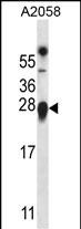
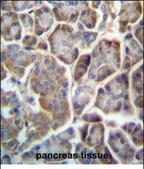
TMED9 Antibody (C-term)
Affinity Purified Rabbit Polyclonal Antibody (Pab)
- SPECIFICATION
- CITATIONS
- PROTOCOLS
- BACKGROUND

Application
| IHC-P, WB, E |
|---|---|
| Primary Accession | Q9BVK6 |
| Other Accession | Q5I0E7, Q99KF1, Q3T133, NP_059980.2 |
| Reactivity | Human |
| Predicted | Bovine, Mouse, Rat |
| Host | Rabbit |
| Clonality | Polyclonal |
| Isotype | Rabbit IgG |
| Calculated MW | 27277 Da |
| Antigen Region | 169-198 aa |
| Gene ID | 54732 |
|---|---|
| Other Names | Transmembrane emp24 domain-containing protein 9, GMP25, Glycoprotein 25L2, p24 family protein alpha-2, p24alpha2, p25, TMED9, GP25L2 |
| Target/Specificity | This TMED9 antibody is generated from rabbits immunized with a KLH conjugated synthetic peptide between 169-198 amino acids from the C-terminal region of human TMED9. |
| Dilution | IHC-P~~1:10~50 WB~~1:1000 E~~Use at an assay dependent concentration. |
| Format | Purified polyclonal antibody supplied in PBS with 0.09% (W/V) sodium azide. This antibody is purified through a protein A column, followed by peptide affinity purification. |
| Storage | Maintain refrigerated at 2-8°C for up to 2 weeks. For long term storage store at -20°C in small aliquots to prevent freeze-thaw cycles. |
| Precautions | TMED9 Antibody (C-term) is for research use only and not for use in diagnostic or therapeutic procedures. |
| Name | TMED9 |
|---|---|
| Synonyms | GP25L2 |
| Function | Appears to be involved in vesicular protein trafficking, mainly in the early secretory pathway. In COPI vesicle-mediated retrograde transport involved in the coatomer recruitment to membranes of the early secretory pathway. Increases coatomer-dependent activity of ARFGAP2. Thought to play a crucial role in the specific retention of p24 complexes in cis-Golgi membranes; specifically contributes to the coupled localization of TMED2 and TMED10 in the cis-Golgi network. May be involved in organization of intracellular membranes, such as of the ER-Golgi intermediate compartment and the Golgi apparatus. Involved in ER localization of PTPN2 isoform PTPB. |
| Cellular Location | Endoplasmic reticulum membrane; Single-pass type I membrane protein. Golgi apparatus, cis-Golgi network membrane; Single-pass type I membrane protein. Endoplasmic reticulum-Golgi intermediate compartment membrane; Single-pass type I membrane protein Golgi apparatus, trans-Golgi network membrane; Single- pass type I membrane protein. Note=Cycles between compartments of the early secretatory pathway |

Thousands of laboratories across the world have published research that depended on the performance of antibodies from Abcepta to advance their research. Check out links to articles that cite our products in major peer-reviewed journals, organized by research category.
info@abcepta.com, and receive a free "I Love Antibodies" mug.

Provided below are standard protocols that you may find useful for product applications.
Background
The specific function of this protein remains unknown.
References
Breuza, L., et al. J. Biol. Chem. 279(45):47242-47253(2004)
Zhang, H., et al. Nat. Biotechnol. 21(6):660-666(2003)
Zhang, H., et al. Nat. Biotechnol. 21(6):660-666(2003)
Renz, M., et al. J. Biol. Chem. 275(14):10429-10436(2000)
Wada, I., et al. J. Biol. Chem. 266(29):19599-19610(1991)
If you have used an Abcepta product and would like to share how it has performed, please click on the "Submit Review" button and provide the requested information. Our staff will examine and post your review and contact you if needed.
If you have any additional inquiries please email technical services at tech@abcepta.com.
Ordering Information
Other Products
Shipping Information
