


 Foundational characteristics of cancer include proliferation, angiogenesis, migration, evasion of apoptosis, and cellular immortality. Find key markers for these cellular processes and antibodies to detect them.
 The SUMOplot™ Analysis Program predicts and scores sumoylation sites in your protein. SUMOylation is a post-translational modification involved in various cellular processes, such as nuclear-cytosolic transport, transcriptional regulation, apoptosis, protein stability, response to stress, and progression through the cell cycle.
 The Autophagy Receptor Motif Plotter predicts and scores autophagy receptor binding sites in your protein. Identifying proteins connected to this pathway is critical to understanding the role of autophagy in physiological as well as pathological processes such as development, differentiation, neurodegenerative diseases, stress, infection, and cancer.
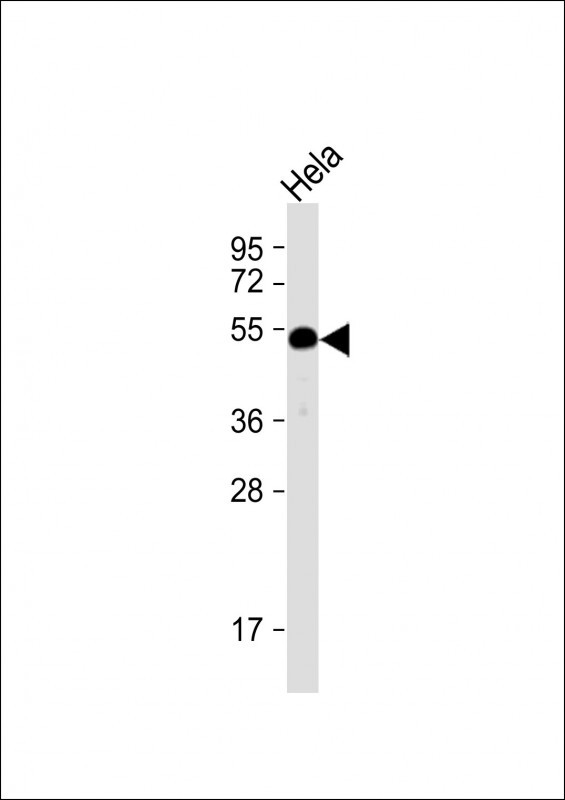
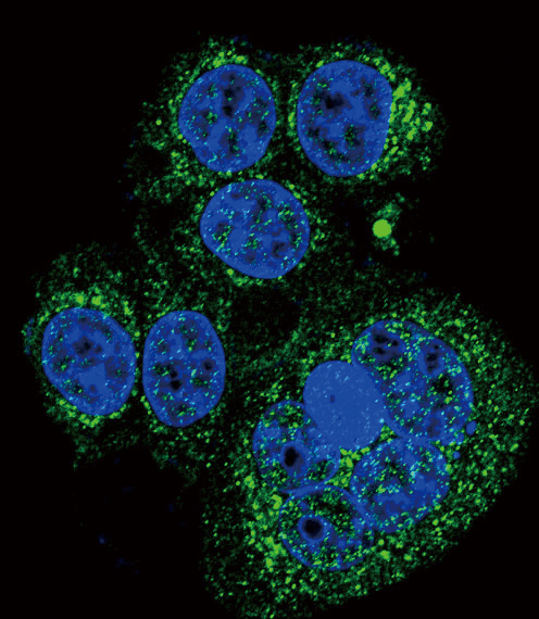
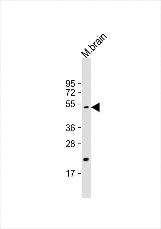
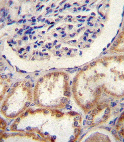
HtrA1 Antibody (N-term)
Purified Rabbit Polyclonal Antibody (Pab)
- SPECIFICATION
- CITATIONS: 2
- PROTOCOLS
- BACKGROUND

Application
| WB, IF, IHC-P, E |
|---|---|
| Primary Accession | Q92743 |
| Reactivity | Human, Mouse |
| Host | Rabbit |
| Clonality | Polyclonal |
| Isotype | Rabbit IgG |
| Calculated MW | 51287 Da |
| Antigen Region | 116-147 aa |
| Gene ID | 5654 |
|---|---|
| Other Names | Serine protease HTRA1, 3421-, High-temperature requirement A serine peptidase 1, L56, Serine protease 11, HTRA1, HTRA, PRSS11 |
| Target/Specificity | This HtrA1 antibody is generated from rabbits immunized with a KLH conjugated synthetic peptide between 116-147 amino acids from the N-terminal region of human HtrA1. |
| Dilution | WB~~1:2000 IF~~1:10~50 IHC-P~~1:10~50 E~~Use at an assay dependent concentration. |
| Format | Purified polyclonal antibody supplied in PBS with 0.09% (W/V) sodium azide. This antibody is prepared by Saturated Ammonium Sulfate (SAS) precipitation followed by dialysis against PBS. |
| Storage | Maintain refrigerated at 2-8°C for up to 2 weeks. For long term storage store at -20°C in small aliquots to prevent freeze-thaw cycles. |
| Precautions | HtrA1 Antibody (N-term) is for research use only and not for use in diagnostic or therapeutic procedures. |
| Name | HTRA1 |
|---|---|
| Synonyms | HTRA, PRSS11 |
| Function | Serine protease with a variety of targets, including extracellular matrix proteins such as fibronectin. HTRA1-generated fibronectin fragments further induce synovial cells to up-regulate MMP1 and MMP3 production. May also degrade proteoglycans, such as aggrecan, decorin and fibromodulin. Through cleavage of proteoglycans, may release soluble FGF-glycosaminoglycan complexes that promote the range and intensity of FGF signals in the extracellular space. Regulates the availability of insulin-like growth factors (IGFs) by cleaving IGF- binding proteins. Inhibits signaling mediated by TGF-beta family members. This activity requires the integrity of the catalytic site, although it is unclear whether TGF-beta proteins are themselves degraded. By acting on TGF-beta signaling, may regulate many physiological processes, including retinal angiogenesis and neuronal survival and maturation during development. Intracellularly, degrades TSC2, leading to the activation of TSC2 downstream targets. |
| Cellular Location | Cell membrane. Secreted Cytoplasm, cytosol. Note=Predominantly secreted (PubMed:15208355). Also found associated with the plasma membrane (PubMed:21297635). |
| Tissue Location | Widely expressed, with strongest expression in placenta (at protein level). Secreted by synovial fibroblasts. Up- regulated in osteoarthritis and rheumatoid arthritis synovial fluids and cartilage as compared with non-arthritic (at protein level) |

Provided below are standard protocols that you may find useful for product applications.
Background
HtrA1 a member of the trypsin family of serine proteases. This protein is a secreted enzyme that is proposed to regulate the availability of insulin-like growth factors (IGFs) by cleaving IGF-binding proteins. It has also been suggested to be a regulator of cell growth.
References
Howes, N., et al., Clin Gastroenterol Hepatol 2(3):252-261 (2004).
Chien, J., et al., Oncogene 23(8):1636-1644 (2004).
Hu, S.I., et al., J. Biol. Chem. 273(51):34406-34412 (1998).
Zumbrunn, J., et al., Genomics 45(2):461-462 (1997).
Zumbrunn, J., et al., FEBS Lett. 398 (2-3), 187-192 (1996).
If you have used an Abcepta product and would like to share how it has performed, please click on the "Submit Review" button and provide the requested information. Our staff will examine and post your review and contact you if needed.
If you have any additional inquiries please email technical services at tech@abcepta.com.
Ordering Information
Other Products
Shipping Information
