


 Foundational characteristics of cancer include proliferation, angiogenesis, migration, evasion of apoptosis, and cellular immortality. Find key markers for these cellular processes and antibodies to detect them.
 The SUMOplot™ Analysis Program predicts and scores sumoylation sites in your protein. SUMOylation is a post-translational modification involved in various cellular processes, such as nuclear-cytosolic transport, transcriptional regulation, apoptosis, protein stability, response to stress, and progression through the cell cycle.
 The Autophagy Receptor Motif Plotter predicts and scores autophagy receptor binding sites in your protein. Identifying proteins connected to this pathway is critical to understanding the role of autophagy in physiological as well as pathological processes such as development, differentiation, neurodegenerative diseases, stress, infection, and cancer.
ANKS1B Antibody (Center)
Affinity Purified Rabbit Polyclonal Antibody (Pab)
- SPECIFICATION
- CITATIONS
- PROTOCOLS
- BACKGROUND

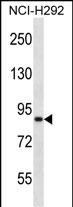
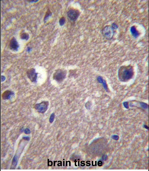
Application
| IHC-P, WB, E |
|---|---|
| Primary Accession | Q7Z6G8 |
| Other Accession | NP_064525.1, NP_690001.3 |
| Reactivity | Human |
| Host | Rabbit |
| Clonality | Polyclonal |
| Isotype | Rabbit IgG |
| Calculated MW | 138066 Da |
| Antigen Region | 570-599 aa |
| Gene ID | 56899 |
|---|---|
| Other Names | Ankyrin repeat and sterile alpha motif domain-containing protein 1B, Amyloid-beta protein intracellular domain-associated protein 1, AIDA-1, E2A-PBX1-associated protein, EB-1, ANKS1B |
| Target/Specificity | This ANKS1B antibody is generated from rabbits immunized with a KLH conjugated synthetic peptide between 570-599 amino acids from the Central region of human ANKS1B. |
| Dilution | IHC-P~~1:10~50 WB~~1:1000 E~~Use at an assay dependent concentration. |
| Format | Purified polyclonal antibody supplied in PBS with 0.09% (W/V) sodium azide. This antibody is purified through a protein A column, followed by peptide affinity purification. |
| Storage | Maintain refrigerated at 2-8°C for up to 2 weeks. For long term storage store at -20°C in small aliquots to prevent freeze-thaw cycles. |
| Precautions | ANKS1B Antibody (Center) is for research use only and not for use in diagnostic or therapeutic procedures. |
| Name | ANKS1B |
|---|---|
| Function | Isoform 2 may participate in the regulation of nucleoplasmic coilin protein interactions in neuronal and transformed cells. Isoform 4 may play a role as a modulator of APP processing. Overexpression can down-regulate APP processing. |
| Cellular Location | Cytoplasm [Isoform 3]: Postsynaptic density. Cell projection, dendritic spine. Nucleus. Nucleus, Cajal body. Note=The synaptic localization requires DLG4 interaction. Translocation to the nucleus in response to stimulation of NMDA receptors (NMDARs) in a calcium-independent manner (By similarity). [Isoform 6]: Nucleus. |
| Tissue Location | Highly expressed in marrow from patients with pre-B ALL associated with the t(1;19) translocation. Strongly expressed in brain and testis. Expressed in fetal brain. Isoform 4 is highly expressed in brain (at protein level). Isoform 6 is expressed in brain and several cancer cell lines. |

Thousands of laboratories across the world have published research that depended on the performance of antibodies from Abcepta to advance their research. Check out links to articles that cite our products in major peer-reviewed journals, organized by research category.
info@abcepta.com, and receive a free "I Love Antibodies" mug.

Provided below are standard protocols that you may find useful for product applications.
Background
Isoform 2 may participate in the regulation of nucleoplasmic coilin protein interactions in neuronal and transformed cells. Isoform 3 can regulate global protein synthesis by altering nucleolar numbers (By similarity). Isoform 4 may play a role as a modulator of APP processing. Overexpression can down-regulate APP processing.
References
Bailey, S.D., et al. Diabetes Care 33(10):2250-2253(2010)
Rose, J.E., et al. Mol. Med. 16 (7-8), 247-253 (2010) :
Pei, D., et al. Genet Test Mol Biomarkers 14(3):433-438(2010)
Wang, M., et al. Stat Biopharm Res 1(4):424-430(2009)
Talmud, P.J., et al. Am. J. Hum. Genet. 85(5):628-642(2009)
If you have used an Abcepta product and would like to share how it has performed, please click on the "Submit Review" button and provide the requested information. Our staff will examine and post your review and contact you if needed.
If you have any additional inquiries please email technical services at tech@abcepta.com.
Ordering Information
Shipping Information
